8A6M | pdb_00008a6m

Phosphatidylserine-dependent synaptic vesicle membrane sculpting by synaptogyrin


Experimental Data Snapshot

  • Method: SOLUTION NMR
  • Conformers Calculated: 470 
  • Conformers Submitted: 10 
  • Selection Criteria: structures with the lowest energy 

wwPDB Validation 3D Report Full Report

Validation slider image for 8A6M

This is version 1.3 of the entry. See complete history

Literature

Phosphatidylserine-dependent structure of synaptogyrin remodels the synaptic vesicle membrane.

Yu, T.Flores-Solis, D.Eastep, G.N.Becker, S.Zweckstetter, M.

(2023) Nat Struct Mol Biol 30: 926-934

  • DOI: https://doi.org/10.1038/s41594-023-01004-9
  • Primary Citation Related Structures: 
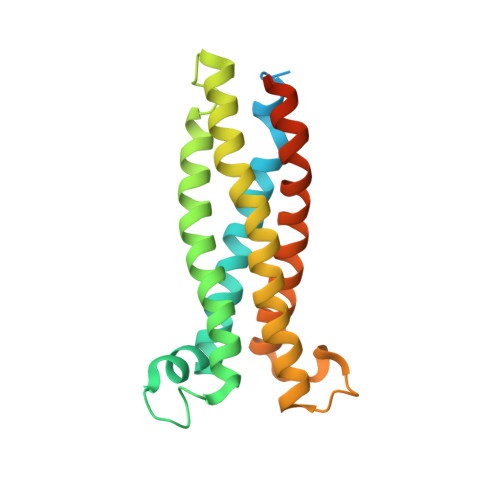
    8A6M

  • PubMed Abstract: 

    Synaptic vesicles are small membrane-enclosed organelles that store neurotransmitters at presynaptic terminals. The uniform morphology of synaptic vesicles is important for brain function, because it enables the storage of well-defined amounts of neurotransmitters and thus reliable synaptic transmission. Here, we show that the synaptic vesicle membrane protein synaptogyrin cooperates with the lipid phosphatidylserine to remodel the synaptic vesicle membrane. Using NMR spectroscopy, we determine the high-resolution structure of synaptogyrin and identify specific binding sites for phosphatidylserine. We further show that phosphatidylserine binding changes the transmembrane structure of synaptogyrin and is critical for membrane bending and the formation of small vesicles. Cooperative binding of phosphatidylserine to both a cytoplasmic and intravesicular lysine-arginine cluster in synaptogyrin is required for the formation of small vesicles. Together with other synaptic vesicle proteins, synaptogyrin thus can sculpt the membrane of synaptic vesicles.


  • Organizational Affiliation
    • German Center for Neurodegenerative Diseases (DZNE), Göttingen, Germany.

Macromolecule Content 

  • Total Structure Weight: 23.36 kDa 
  • Atom Count: 1,276 
  • Modeled Residue Count: 161 
  • Deposited Residue Count: 210 
  • Unique protein chains: 1

Macromolecules

Find similar proteins by:|  3D Structure
Entity ID: 1
MoleculeChains  Sequence LengthOrganismDetailsImage
Isoform 1B of Synaptogyrin-1210Homo sapiensMutation(s): 0 
Gene Names: SYNGR1
Membrane Entity: Yes 
UniProt & NIH Common Fund Data Resources
Find proteins for O43759 (Homo sapiens)
Explore O43759 
Go to UniProtKB:  O43759
PHAROS:  O43759
GTEx:  ENSG00000100321 
Entity Groups
Sequence Clusters30% Identity50% Identity70% Identity90% Identity95% Identity100% Identity
UniProt GroupO43759
Sequence Annotations
Expand
Reference Sequence

Experimental Data & Validation

Experimental Data

  • Method: SOLUTION NMR
  • Conformers Calculated: 470 
  • Conformers Submitted: 10 
  • Selection Criteria: structures with the lowest energy 

Structure Validation

View Full Validation Report


Entry History 

& Funding Information

Deposition Data


Funding OrganizationLocationGrant Number
German Research Foundation (DFG)Germany105286809

Revision History  (Full details and data files)

  • Version 1.0: 2023-05-24
    Type: Initial release
  • Version 1.1: 2023-05-31
    Changes: Database references
  • Version 1.2: 2023-07-26
    Changes: Database references
  • Version 1.3: 2024-06-19
    Changes: Data collection, Database references