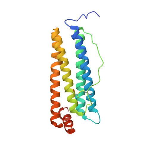
8A5N

X-ray structure of human H-chain ferritin treated with SDS


Experimental Data Snapshot

  • Method: X-RAY DIFFRACTION
  • Resolution: 1.52 Å
  • R-Value Free: 0.190 
  • R-Value Work: 0.151 

Starting Model: experimental
View more details

wwPDB Validation   3D Report Full Report


This is version 1.2 of the entry. See complete history


Literature

A new and efficient procedure to load bioactive molecules within the human heavy-chain ferritin nanocage.

Lucignano, R.Stanzione, I.Ferraro, G.Di Girolamo, R.Cane, C.Di Somma, A.Duilio, A.Merlino, A.Picone, D.

(2023) Front Mol Biosci 10: 1008985-1008985

  • DOI: https://doi.org/10.3389/fmolb.2023.1008985
  • Primary Citation of Related Structures:  
    8A2L, 8A2M, 8A5N

  • PubMed Abstract: 

    For their easy and high-yield recombinant production, their high stability in a wide range of physico-chemical conditions and their characteristic hollow structure, ferritins (Fts) are considered useful scaffolds to encapsulate bioactive molecules. Notably, for the absence of immunogenicity and the selective interaction with tumor cells, the nanocages constituted by the heavy chain of the human variant of ferritin (hHFt) are optimal candidates for the delivery of anti-cancer drugs. hHFt nanocages can be disassembled and reassembled in vitro to allow the loading of cargo molecules, however the currently available protocols present some relevant drawbacks. Indeed, protein disassembly is achieved by exposure to extreme pH (either acidic or alkaline), followed by incubation at neutral pH to allow reassembly, but the final protein recovery and homogeneity are not satisfactory. Moreover, the exposure to extreme pH may affect the structure of the molecule to be loaded. In this paper, we report an alternative, efficient and reproducible procedure to reversibly disassemble hHFt under mild pH conditions. We demonstrate that a small amount of sodium dodecyl sulfate (SDS) is sufficient to disassemble the nanocage, which quantitatively reassembles upon SDS removal. Electron microscopy and X-ray crystallography show that the reassembled protein is identical to the untreated one. The newly developed procedure was used to encapsulate two small molecules. When compared to the existing disassembly/reassembly procedures, our approach can be applied in a wide range of pH values and temperatures, is compatible with a larger number of cargos and allows a higher protein recovery.


  • Organizational Affiliation

    Department of Chemical Sciences, University of Naples Federico II, Naples, Italy.


Macromolecules
Find similar proteins by:  (by identity cutoff)  |  3D Structure
Entity ID: 1
MoleculeChains Sequence LengthOrganismDetailsImage
Ferritin heavy chainA [auth AAA]182Homo sapiensMutation(s): 0 
Gene Names: FTH1FTHFTHL6OK/SW-cl.84PIG15
EC: 1.16.3.1
UniProt & NIH Common Fund Data Resources
Find proteins for P02794 (Homo sapiens)
Explore P02794 
Go to UniProtKB:  P02794
PHAROS:  P02794
GTEx:  ENSG00000167996 
Entity Groups  
Sequence Clusters30% Identity50% Identity70% Identity90% Identity95% Identity100% Identity
UniProt GroupP02794
Sequence Annotations
Expand
  • Reference Sequence
Small Molecules
Ligands 4 Unique
IDChains Name / Formula / InChI Key2D Diagram3D Interactions
GOL
Query on GOL

Download Ideal Coordinates CCD File 
D [auth AAA]GLYCEROL
C3 H8 O3
PEDCQBHIVMGVHV-UHFFFAOYSA-N
FE
Query on FE

Download Ideal Coordinates CCD File 
M [auth AAA]FE (III) ION
Fe
VTLYFUHAOXGGBS-UHFFFAOYSA-N
CL
Query on CL

Download Ideal Coordinates CCD File 
B [auth AAA]
C [auth AAA]
E [auth AAA]
F [auth AAA]
G [auth AAA]
B [auth AAA],
C [auth AAA],
E [auth AAA],
F [auth AAA],
G [auth AAA],
H [auth AAA],
I [auth AAA],
J [auth AAA],
K [auth AAA],
L [auth AAA]
CHLORIDE ION
Cl
VEXZGXHMUGYJMC-UHFFFAOYSA-M
MG
Query on MG

Download Ideal Coordinates CCD File 
N [auth AAA]
O [auth AAA]
P [auth AAA]
Q [auth AAA]
R [auth AAA]
N [auth AAA],
O [auth AAA],
P [auth AAA],
Q [auth AAA],
R [auth AAA],
S [auth AAA],
T [auth AAA],
U [auth AAA],
V [auth AAA],
W [auth AAA]
MAGNESIUM ION
Mg
JLVVSXFLKOJNIY-UHFFFAOYSA-N
Experimental Data & Validation

Experimental Data

  • Method: X-RAY DIFFRACTION
  • Resolution: 1.52 Å
  • R-Value Free: 0.190 
  • R-Value Work: 0.151 
  • Space Group: F 4 3 2
Unit Cell:
Length ( Å )Angle ( ˚ )
a = 183.92α = 90
b = 183.92β = 90
c = 183.92γ = 90
Software Package:
Software NamePurpose
REFMACrefinement
autoPROCdata reduction
autoPROCdata scaling
PHASERphasing

Structure Validation

View Full Validation Report


Entry History & Funding Information

Deposition Data


Funding OrganizationLocationGrant Number
Not funded--

Revision History  (Full details and data files)

  • Version 1.0: 2023-02-08
    Type: Initial release
  • Version 1.1: 2024-02-07
    Changes: Data collection, Refinement description
  • Version 1.2: 2024-10-09
    Changes: Structure summary