8A49 | pdb_00008a49

Endoglycosidase S in complex with IgG1 Fc


Experimental Data Snapshot

  • Method: X-RAY DIFFRACTION
  • Resolution: 3.45 Å
  • R-Value Free: 
    0.311 (Depositor), 0.315 (DCC) 
  • R-Value Work: 
    0.253 (Depositor), 0.258 (DCC) 

Starting Model: experimental
View more details

wwPDB Validation 3D Report Full Report

Validation slider image for 8A49

This is version 1.4 of the entry. See complete history

Literature

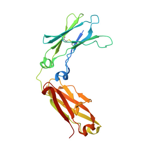
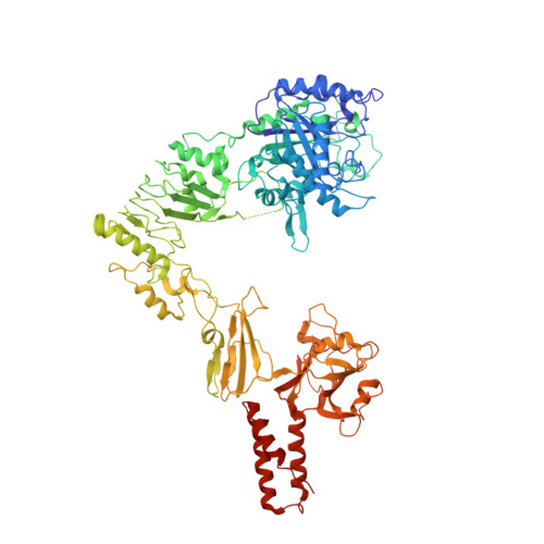
Extensive substrate recognition by the streptococcal antibody-degrading enzymes IdeS and EndoS.

Sudol, A.S.L.Butler, J.Ivory, D.P.Tews, I.Crispin, M.

(2022) Nat Commun 13: 7801-7801

  • DOI: https://doi.org/10.1038/s41467-022-35340-z
  • Primary Citation Related Structures: 
    8A47, 8A48, 8A49

  • PubMed Abstract: 

    Enzymatic cleavage of IgG antibodies is a common strategy used by pathogenic bacteria to ablate immune effector function. The Streptococcus pyogenes bacterium secretes the protease IdeS and the glycosidase EndoS, which specifically catalyse cleavage and deglycosylation of human IgG, respectively. IdeS has received clinical approval for kidney transplantation in hypersensitised individuals, while EndoS has found application in engineering antibody glycosylation. We present crystal structures of both enzymes in complex with their IgG1 Fc substrate, which was achieved using Fc engineering to disfavour preferential Fc crystallisation. The IdeS protease displays extensive Fc recognition and encases the antibody hinge. Conversely, the glycan hydrolase domain in EndoS traps the Fc glycan in a "flipped-out" conformation, while additional recognition of the Fc peptide is driven by the so-called carbohydrate binding module. In this work, we reveal the molecular basis of antibody recognition by bacterial enzymes, providing a template for the development of next-generation enzymes.


  • Organizational Affiliation
    • School of Biological Sciences, University of Southampton, Southampton, SO17 1BJ, UK.

Macromolecule Content 

  • Total Structure Weight: 257.49 kDa 
  • Atom Count: 17,087 
  • Modeled Residue Count: 2,177 
  • Deposited Residue Count: 2,266 
  • Unique protein chains: 2

Macromolecules

Find similar proteins by:|  3D Structure
Entity ID: 1
MoleculeChains  Sequence LengthOrganismDetailsImage
IgG1 FcA,
D [auth B]
227Homo sapiensMutation(s): 0 
UniProt
Find proteins for P0DOX5 (Homo sapiens)
Explore P0DOX5 
Go to UniProtKB:  P0DOX5
Entity Groups
Sequence Clusters30% Identity50% Identity70% Identity90% Identity95% Identity100% Identity
UniProt GroupP0DOX5
Glycosylation
Glycosylation Sites: 1
Sequence Annotations
Expand
Reference Sequence
Find similar proteins by:|  3D Structure
Entity ID: 2
MoleculeChains  Sequence LengthOrganismDetailsImage
Secreted endoglycosidase EndoSB [auth C],
C [auth D]
906Streptococcus pyogenesMutation(s): 2 
Gene Names: ndoS
EC: 3.2.1.96
UniProt
Find proteins for Q99Y92 (Streptococcus pyogenes serotype M1)
Explore Q99Y92 
Go to UniProtKB:  Q99Y92
Entity Groups
Sequence Clusters30% Identity50% Identity70% Identity90% Identity95% Identity100% Identity
UniProt GroupQ99Y92
Sequence Annotations
Expand
Reference Sequence

Oligosaccharides

Help  
Entity ID: 3
MoleculeChains Length2D Diagram GlycosylationD Interactions
2-acetamido-2-deoxy-beta-D-glucopyranose-(1-2)-alpha-D-mannopyranose-(1-3)-[2-acetamido-2-deoxy-beta-D-glucopyranose-(1-2)-alpha-D-mannopyranose-(1-6)]beta-D-mannopyranose-(1-4)-2-acetamido-2-deoxy-beta-D-glucopyranose-(1-4)-[alpha-L-fucopyranose-(1-6)]2-acetamido-2-deoxy-beta-D-glucopyranose
E
8N-Glycosylation
Glycosylation Resources
GlyTouCan: G80858MF
GlyCosmos: G80858MF
GlyGen: G80858MF

Experimental Data & Validation

Experimental Data

  • Method: X-RAY DIFFRACTION
  • Resolution: 3.45 Å
  • R-Value Free:  0.311 (Depositor), 0.315 (DCC) 
  • R-Value Work:  0.253 (Depositor), 0.258 (DCC) 
Space Group: P 21 21 21
Unit Cell:
Length ( Å )Angle ( ˚ )
a = 96.529α = 90
b = 174.294β = 90
c = 193.059γ = 90
Software Package:
Software NamePurpose
REFMACrefinement
DIALSdata reduction
DIALSdata scaling
MOLREPphasing

Structure Validation

View Full Validation Report


Entry History 

& Funding Information

Deposition Data


Funding OrganizationLocationGrant Number
Other private--

Revision History  (Full details and data files)

  • Version 1.0: 2022-11-30
    Type: Initial release
  • Version 1.1: 2022-12-28
    Changes: Database references
  • Version 1.2: 2023-01-11
    Changes: Database references
  • Version 1.3: 2024-01-31
    Changes: Data collection, Refinement description
  • Version 1.4: 2024-11-20
    Changes: Structure summary