8A3B | pdb_00008a3b

Cryo-EM structure of mouse Pannexin 1 purified in Salipro nanoparticles


Experimental Data Snapshot

  • Method: ELECTRON MICROSCOPY
  • Resolution: 3.10 Å
  • Aggregation State: PARTICLE 
  • Reconstruction Method: SINGLE PARTICLE 

wwPDB Validation 3D Report Full Report

Validation slider image for 8A3B

This is version 1.1 of the entry. See complete history

Literature

Direct cell extraction of membrane proteins for structure-function analysis.

Drulyte, I.Gutgsell, A.R.Lloris-Garcera, P.Liss, M.Geschwindner, S.Radjainia, M.Frauenfeld, J.Loving, R.

(2023) Sci Rep 13: 1420-1420

  • DOI: https://doi.org/10.1038/s41598-023-28455-w
  • Primary Citation Related Structures: 
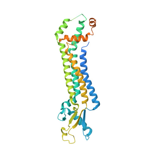
    8A3B

  • PubMed Abstract: 

    Membrane proteins are the largest group of therapeutic targets in a variety of disease areas and yet, they remain particularly difficult to investigate. We have developed a novel one-step approach for the incorporation of membrane proteins directly from cells into lipid Salipro nanoparticles. Here, with the pannexin1 channel as a case study, we demonstrate the applicability of this method for structure-function analysis using SPR and cryo-EM.


  • Organizational Affiliation
    • Thermo Fisher Scientific, Materials and Structural Analysis, Achtseweg Noord 5, 5651 GG, Eindhoven, The Netherlands.

Macromolecule Content 

  • Total Structure Weight: 353.33 kDa 
  • Atom Count: 14,448 
  • Modeled Residue Count: 1,792 
  • Deposited Residue Count: 3,129 
  • Unique protein chains: 1

Macromolecules

Find similar proteins by:|  3D Structure
Entity ID: 1
MoleculeChains  Sequence LengthOrganismDetailsImage
Pannexin-1
A, B, C, D, E
A, B, C, D, E, F, G
447Mus musculusMutation(s): 0 
Gene Names: Panx1
Membrane Entity: Yes 
UniProt & NIH Common Fund Data Resources
Find proteins for Q9JIP4 (Mus musculus)
Explore Q9JIP4 
Go to UniProtKB:  Q9JIP4
IMPC:  MGI:1860055
Entity Groups
Sequence Clusters30% Identity50% Identity70% Identity90% Identity95% Identity100% Identity
UniProt GroupQ9JIP4
Sequence Annotations
Expand
Reference Sequence

Experimental Data & Validation

Experimental Data

  • Method: ELECTRON MICROSCOPY
  • Resolution: 3.10 Å
  • Aggregation State: PARTICLE 
  • Reconstruction Method: SINGLE PARTICLE 
EM Software:
TaskSoftware PackageVersion
RECONSTRUCTIONREFMAC5.8
MODEL REFINEMENTISOLDE1.3
MODEL REFINEMENTREFMAC5.8

Structure Validation

View Full Validation Report


Entry History 

& Funding Information

Deposition Data

  • Released Date: 2023-02-08 
  • Deposition Author(s): Drulyte, I.

Funding OrganizationLocationGrant Number
Not funded--

Revision History  (Full details and data files)

  • Version 1.0: 2023-02-08
    Type: Initial release
  • Version 1.1: 2024-07-24
    Changes: Data collection