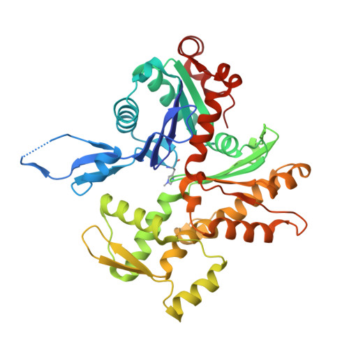
Structural basis of actin filament assembly and aging.
Oosterheert, W., Klink, B.U., Belyy, A., Pospich, S., Raunser, S.(2022) Nature 611: 374-379
- PubMed: 36289337
- DOI: https://doi.org/10.1038/s41586-022-05241-8
- Primary Citation of Related Structures:
8A2R, 8A2S, 8A2T, 8A2U, 8A2Y, 8A2Z - PubMed Abstract:
The dynamic turnover of actin filaments (F-actin) controls cellular motility in eukaryotes and is coupled to changes in the F-actin nucleotide state 1-3 . It remains unclear how F-actin hydrolyses ATP and subsequently undergoes subtle conformational rearrangements that ultimately lead to filament depolymerization by actin-binding proteins. Here we present cryo-electron microscopy structures of F-actin in all nucleotide states, polymerized in the presence of Mg 2+ or Ca 2+ at approximately 2.2 Å resolution. The structures show that actin polymerization induces the relocation of water molecules in the nucleotide-binding pocket, activating one of them for the nucleophilic attack of ATP. Unexpectedly, the back door for the subsequent release of inorganic phosphate (P i ) is closed in all structures, indicating that P i release occurs transiently. The small changes in the nucleotide-binding pocket after ATP hydrolysis and P i release are sensed by a key amino acid, amplified and transmitted to the filament periphery. Furthermore, differences in the positions of water molecules in the nucleotide-binding pocket explain why Ca 2+ -actin shows slower polymerization rates than Mg 2+ -actin. Our work elucidates the solvent-driven rearrangements that govern actin filament assembly and aging and lays the foundation for the rational design of drugs and small molecules for imaging and therapeutic applications.
- Department of Structural Biochemistry, Max Planck Institute of Molecular Physiology, Dortmund, Germany.
Organizational Affiliation:


