7ZUN

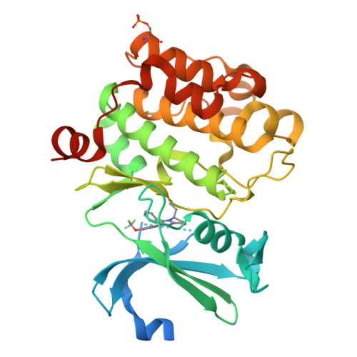
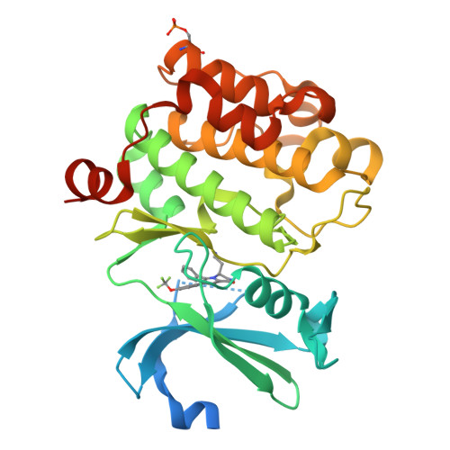
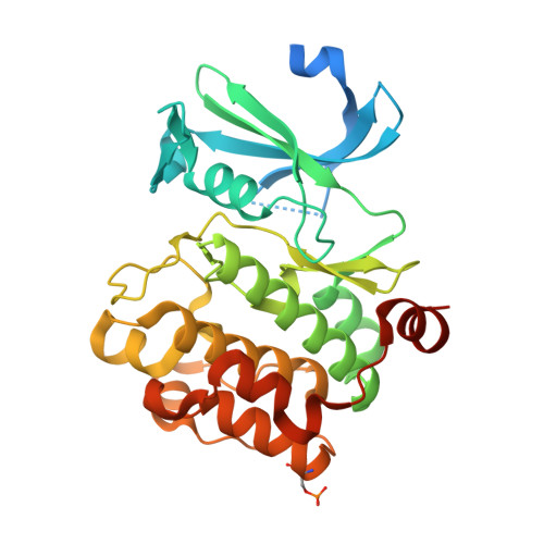
Crystal structure of PIM1 in complex with a Pyrrolo-Pyrazinone compound


Experimental Data Snapshot

  • Method: X-RAY DIFFRACTION
  • Resolution: 2.50 Å
  • R-Value Free: 0.235 
  • R-Value Work: 0.174 

Starting Model: experimental
View more details

wwPDB Validation   3D Report Full Report


Ligand Structure Quality Assessment 


This is version 2.2 of the entry. See complete history


Literature

Stereoselective synthesis of 3,4-dihydropyrrolo[1,2-a]pyrazin-1(2H)-one derivatives as PIM kinase inhibitors inspired from marine alkaloids.

Casuscelli, F.Ardini, E.Avanzi, N.Badari, A.Casale, E.Disingrini, T.Donati, D.Ermoli, A.Felder, E.R.Galvani, A.Isacchi, A.Menichincheri, M.Montemartini, M.Orrenius, C.Piutti, C.Salom, B.Papeo, G.

(2022) Chirality 34: 1437-1452

  • DOI: https://doi.org/10.1002/chir.23501
  • Primary Citation of Related Structures:  
    7ZUN

  • PubMed Abstract: 

    We previously demonstrated that natural product-inspired 3,4-dihydropyrrolo[1,2-a]pyrazin-1(2H)-ones derivatives delivered potent and selective PIM kinases inhibitors however with non-optimal ADME/PK properties and modest oral bioavailability. Herein, we describe a structure-based scaffold decoration and a stereoselective approach to this chemical class. The synthesis, structure-activity relationship studies, chiral analysis, and pharmacokinetic data of compounds from this inhibitor class are presented herein. Compound 20c demonstrated excellent potency on PIM1 and PIM2 with exquisite kinases selectivity and PK properties that efficiently and dose-dependently promoted c-Myc degradation and appear to be promising lead compounds for further development.


  • Organizational Affiliation

    Oncology, Nerviano Medical Sciences, Nerviano (Mi), Italy.


Macromolecules
Find similar proteins by:  (by identity cutoff)  |  3D Structure
Entity ID: 1
MoleculeChains Sequence LengthOrganismDetailsImage
Isoform 2 of Serine/threonine-protein kinase pim-1314Homo sapiensMutation(s): 0 
Gene Names: PIM1
EC: 2.7.11.1
UniProt & NIH Common Fund Data Resources
Find proteins for P11309 (Homo sapiens)
Explore P11309 
Go to UniProtKB:  P11309
PHAROS:  P11309
GTEx:  ENSG00000137193 
Entity Groups  
Sequence Clusters30% Identity50% Identity70% Identity90% Identity95% Identity100% Identity
UniProt GroupP11309
Sequence Annotations
Expand
  • Reference Sequence
Small Molecules
Ligands 1 Unique
IDChains Name / Formula / InChI Key2D Diagram3D Interactions
JYO (Subject of Investigation/LOI)
Query on JYO

Download Ideal Coordinates CCD File 
B [auth A](4~{S})-4-(2-azanylethyl)-6-phenyl-7-[3-(trifluoromethyloxy)phenyl]-3,4-dihydropyrrolo[1,2-a]pyrazin-1-ol
C22 H20 F3 N3 O2
WTGCKCDNZFLRCX-INIZCTEOSA-N
Modified Residues  1 Unique
IDChains TypeFormula2D DiagramParent
SEP
Query on SEP
A
L-PEPTIDE LINKINGC3 H8 N O6 PSER
Experimental Data & Validation

Experimental Data

  • Method: X-RAY DIFFRACTION
  • Resolution: 2.50 Å
  • R-Value Free: 0.235 
  • R-Value Work: 0.174 
  • Space Group: P 65
Unit Cell:
Length ( Å )Angle ( ˚ )
a = 95.09α = 90
b = 95.09β = 90
c = 80.181γ = 120
Software Package:
Software NamePurpose
REFMACrefinement
XDSdata reduction
Aimlessdata scaling
REFMACphasing

Structure Validation

View Full Validation Report


Ligand Structure Quality Assessment 


Entry History & Funding Information

Deposition Data

  • Released Date: 2022-10-05 
  • Deposition Author(s): Casale, E.

Funding OrganizationLocationGrant Number
Not funded--

Revision History  (Full details and data files)

  • Version 1.0: 2022-10-05
    Type: Initial release
  • Version 2.0: 2022-10-12
    Changes: Atomic model, Database references
  • Version 2.1: 2024-01-31
    Changes: Data collection, Refinement description
  • Version 2.2: 2024-11-06
    Changes: Structure summary