7ZUB | pdb_00007zub

Cryo-EM structure of the indirubin-bound Hsp90-XAP2-AHR complex


Experimental Data Snapshot

  • Method: ELECTRON MICROSCOPY
  • Resolution: 2.85 Å
  • Aggregation State: PARTICLE 
  • Reconstruction Method: SINGLE PARTICLE 

Starting Models: experimental
View more details

wwPDB Validation 3D Report Full Report

Validation slider image for 7ZUB

This is version 1.3 of the entry. See complete history

Literature

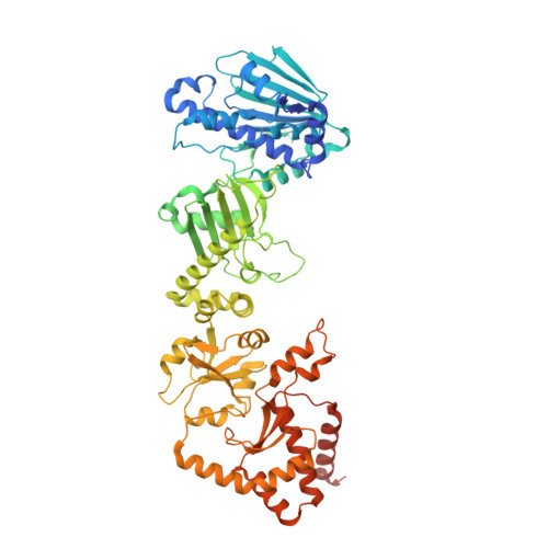
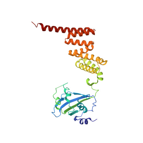
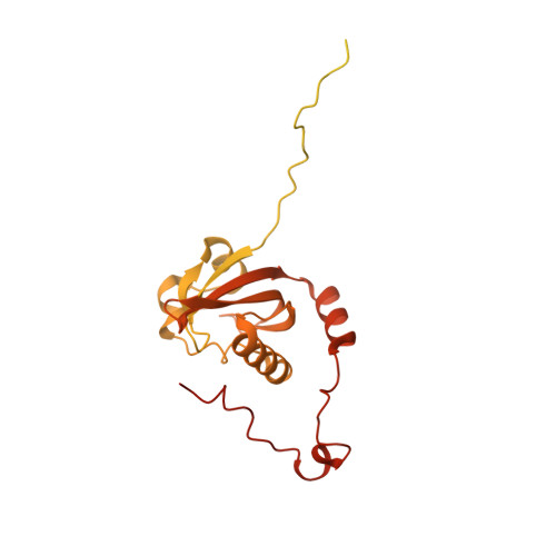
Cryo-EM structure of the agonist-bound Hsp90-XAP2-AHR cytosolic complex.

Gruszczyk, J.Grandvuillemin, L.Lai-Kee-Him, J.Paloni, M.Savva, C.G.Germain, P.Grimaldi, M.Boulahtouf, A.Kwong, H.S.Bous, J.Ancelin, A.Bechara, C.Barducci, A.Balaguer, P.Bourguet, W.

(2022) Nat Commun 13: 7010-7010

  • DOI: https://doi.org/10.1038/s41467-022-34773-w
  • Primary Citation Related Structures: 
    7ZUB

  • PubMed Abstract: 

    The aryl hydrocarbon receptor (AHR) is a ligand-dependent transcription factor that mediates a broad spectrum of (patho)physiological processes in response to numerous substances including pollutants, natural products and metabolites. However, the scarcity of structural data precludes understanding of how AHR is activated by such diverse compounds. Our 2.85 Å structure of the human indirubin-bound AHR complex with the chaperone Hsp90 and the co-chaperone XAP2, reported herein, reveals a closed conformation Hsp90 dimer with AHR threaded through its lumen and XAP2 serving as a brace. Importantly, we disclose the long-awaited structure of the AHR PAS-B domain revealing a unique organisation of the ligand-binding pocket and the structural determinants of ligand-binding specificity and promiscuity of the receptor. By providing structural details of the molecular initiating event leading to AHR activation, our study rationalises almost forty years of biochemical data and provides a framework for future mechanistic studies and structure-guided drug design.


  • Organizational Affiliation
    • CBS (Centre de Biologie Structurale), Univ Montpellier, CNRS, Inserm, Montpellier, France. jakub.gruszczyk@cbs.cnrs.fr.

Macromolecule Content 

  • Total Structure Weight: 257.37 kDa 
  • Atom Count: 14,047 
  • Modeled Residue Count: 1,722 
  • Deposited Residue Count: 2,231 
  • Unique protein chains: 3

Macromolecules

Find similar proteins by:|  3D Structure
Entity ID: 1
MoleculeChains  Sequence LengthOrganismDetailsImage
Heat shock protein HSP 90-beta
A, B
730Homo sapiensMutation(s): 0 
Gene Names: HSP90AB1HSP90BHSPC2HSPCB
UniProt & NIH Common Fund Data Resources
Find proteins for P08238 (Homo sapiens)
Explore P08238 
Go to UniProtKB:  P08238
PHAROS:  P08238
GTEx:  ENSG00000096384 
Entity Groups
Sequence Clusters30% Identity50% Identity70% Identity90% Identity95% Identity100% Identity
UniProt GroupP08238
Sequence Annotations
Expand
Reference Sequence
Find similar proteins by:|  3D Structure
Entity ID: 2
MoleculeChains  Sequence LengthOrganismDetailsImage
AH receptor-interacting protein330Homo sapiensMutation(s): 0 
Gene Names: AIPXAP2
UniProt & NIH Common Fund Data Resources
Find proteins for O00170 (Homo sapiens)
Explore O00170 
Go to UniProtKB:  O00170
PHAROS:  O00170
GTEx:  ENSG00000110711 
Entity Groups
Sequence Clusters30% Identity50% Identity70% Identity90% Identity95% Identity100% Identity
UniProt GroupO00170
Sequence Annotations
Expand
Reference Sequence
Find similar proteins by:|  3D Structure
Entity ID: 3
MoleculeChains  Sequence LengthOrganismDetailsImage
Aryl hydrocarbon receptor441Homo sapiensMutation(s): 0 
Gene Names: AHRBHLHE76
UniProt & NIH Common Fund Data Resources
Find proteins for P35869 (Homo sapiens)
Explore P35869 
Go to UniProtKB:  P35869
PHAROS:  P35869
GTEx:  ENSG00000106546 
Entity Groups
Sequence Clusters30% Identity50% Identity70% Identity90% Identity95% Identity100% Identity
UniProt GroupP35869
Sequence Annotations
Expand
Reference Sequence

Small Molecules

Ligands 4 Unique
IDChains Name / Formula / InChI Key2D Diagram3D Interactions
ADP

Query on ADP


Download:Ideal Coordinates CCD File
E [auth A],
H [auth B]
ADENOSINE-5'-DIPHOSPHATE
C10 H15 N5 O10 P2
XTWYTFMLZFPYCI-KQYNXXCUSA-N
JY6
(Subject of Investigation/LOI)

Query on JY6


Download:Ideal Coordinates CCD File
K [auth D](3~{Z})-3-(3-oxidanylidene-1~{H}-indol-2-ylidene)-1~{H}-indol-2-one
C16 H10 N2 O2
CRDNMYFJWFXOCH-YPKPFQOOSA-N
MOO

Query on MOO


Download:Ideal Coordinates CCD File
G [auth A],
J [auth B]
MOLYBDATE ION
Mo O4
MEFBJEMVZONFCJ-UHFFFAOYSA-N
MG

Query on MG


Download:Ideal Coordinates CCD File
F [auth A],
I [auth B]
MAGNESIUM ION
Mg
JLVVSXFLKOJNIY-UHFFFAOYSA-N
Binding Affinity Annotations 
IDSourceBinding Affinity
JY6 BindingDB:  7ZUB EC50: 0.2 (nM) from 1 assay(s)

Experimental Data & Validation

Experimental Data

  • Method: ELECTRON MICROSCOPY
  • Resolution: 2.85 Å
  • Aggregation State: PARTICLE 
  • Reconstruction Method: SINGLE PARTICLE 
EM Software:
TaskSoftware PackageVersion
RECONSTRUCTIONRELION3.1.1
MODEL REFINEMENTPHENIX1.20

Structure Validation

View Full Validation Report


Entry History 

& Funding Information

Deposition Data


Funding OrganizationLocationGrant Number
European Union (EU)European Union825489
ATIP-AvenirFranceR20059SP
Montpellier University of Excellence (MUSE)FranceANR-16-IDEX-0006

Revision History  (Full details and data files)

  • Version 1.0: 2022-11-23
    Type: Initial release
  • Version 1.1: 2022-11-30
    Changes: Data collection, Database references
  • Version 1.2: 2022-12-07
    Changes: Database references
  • Version 1.3: 2024-07-24
    Changes: Data collection, Refinement description