7ZSC | pdb_00007zsc

Crystal structure of the heterodimeric human C-P4H-II with truncated alpha subunit (C-P4H-II delta281)


Experimental Data Snapshot

  • Method: X-RAY DIFFRACTION
  • Resolution: 3.85 Å
  • R-Value Free: 
    0.278 (Depositor), 0.299 (DCC) 
  • R-Value Work: 
    0.244 (Depositor), 0.261 (DCC) 
  • R-Value Observed: 
    0.247 (Depositor) 

Starting Models: experimental
View more details

wwPDB Validation 3D Report Full Report

Validation slider image for 7ZSC

This is version 1.3 of the entry. See complete history

Literature

Crystal structure of the collagen prolyl 4-hydroxylase (C-P4H) catalytic domain complexed with PDI: Toward a model of the C-P4H alpha 2 beta 2 tetramer.

Murthy, A.V.Sulu, R.Lebedev, A.Salo, A.M.Korhonen, K.Venkatesan, R.Tu, H.Bergmann, U.Janis, J.Laitaoja, M.Ruddock, L.W.Myllyharju, J.Koski, M.K.Wierenga, R.K.

(2022) J Biological Chem 298: 102614-102614

  • DOI: https://doi.org/10.1016/j.jbc.2022.102614
  • Primary Citation Related Structures: 
    7ZSC

  • PubMed Abstract: 

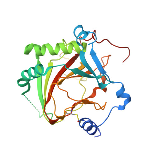
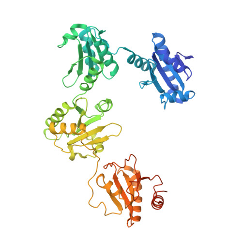
    Collagen prolyl 4-hydroxylases (C-P4H) are α 2 β 2 tetramers, which catalyze the prolyl 4-hydroxylation of procollagen, allowing for the formation of the stable triple-helical collagen structure in the endoplasmic reticulum. The C-P4H α-subunit provides the N-terminal dimerization domain, the middle peptide-substrate-binding (PSB) domain, and the C-terminal catalytic (CAT) domain, whereas the β-subunit is identical to the enzyme protein disulfide isomerase (PDI). The structure of the N-terminal part of the α-subunit (N-terminal region and PSB domain) is known, but the structures of the PSB-CAT linker region and the CAT domain as well as its mode of assembly with the β/PDI subunit, are unknown. Here, we report the crystal structure of the CAT domain of human C-P4H-II complexed with the intact β/PDI subunit, at 3.8 Å resolution. The CAT domain interacts with the a, b', and a' domains of the β/PDI subunit, such that the CAT active site is facing bulk solvent. The structure also shows that the C-P4H-II CAT domain has a unique N-terminal extension, consisting of α-helices and a β-strand, which is the edge strand of its major antiparallel β-sheet. This extra region of the CAT domain interacts tightly with the β/PDI subunit, showing that the CAT-PDI interface includes an intersubunit disulfide bridge with the a' domain and tight hydrophobic interactions with the b' domain. Using this new information, the structure of the mature C-P4H-II α 2 β 2 tetramer is predicted. The model suggests that the CAT active-site properties are modulated by α-helices of the N-terminal dimerization domains of both subunits of the α 2 -dimer.


  • Organizational Affiliation
    • Faculty of Biochemistry and Molecular Medicine, University of Oulu, Oulu, Finland; Biocenter Oulu, University of Oulu, Oulu, Finland.

Macromolecule Content 

  • Total Structure Weight: 171.76 kDa 
  • Atom Count: 11,098 
  • Modeled Residue Count: 1,384 
  • Deposited Residue Count: 1,508 
  • Unique protein chains: 2

Macromolecules

Find similar proteins by:|  3D Structure
Entity ID: 1
MoleculeChains  Sequence LengthOrganismDetailsImage
Prolyl 4-hydroxylase subunit alpha-2
A, B
262Homo sapiensMutation(s): 0 
Gene Names: P4HA2UNQ290/PRO330
EC: 1.14.11.2
UniProt & NIH Common Fund Data Resources
Find proteins for O15460 (Homo sapiens)
Explore O15460 
Go to UniProtKB:  O15460
PHAROS:  O15460
GTEx:  ENSG00000072682 
Entity Groups
Sequence Clusters30% Identity50% Identity70% Identity90% Identity95% Identity100% Identity
UniProt GroupO15460
Sequence Annotations
Expand
Reference Sequence
Find similar proteins by:|  3D Structure
Entity ID: 2
MoleculeChains  Sequence LengthOrganismDetailsImage
Protein disulfide-isomerase
C, D
492Homo sapiensMutation(s): 0 
Gene Names: P4HBERBA2LPDIPDIA1PO4DB
EC: 5.3.4.1
UniProt & NIH Common Fund Data Resources
Find proteins for P07237 (Homo sapiens)
Explore P07237 
Go to UniProtKB:  P07237
PHAROS:  P07237
GTEx:  ENSG00000185624 
Entity Groups
Sequence Clusters30% Identity50% Identity70% Identity90% Identity95% Identity100% Identity
UniProt GroupP07237
Sequence Annotations
Expand
Reference Sequence

Experimental Data & Validation

Experimental Data

  • Method: X-RAY DIFFRACTION
  • Resolution: 3.85 Å
  • R-Value Free:  0.278 (Depositor), 0.299 (DCC) 
  • R-Value Work:  0.244 (Depositor), 0.261 (DCC) 
  • R-Value Observed: 0.247 (Depositor) 
Space Group: H 3
Unit Cell:
Length ( Å )Angle ( ˚ )
a = 252.879α = 90
b = 252.879β = 90
c = 89.4γ = 120
Software Package:
Software NamePurpose
BUSTERrefinement
XDSdata reduction
Aimlessdata scaling
MOLREPphasing
PHASERphasing

Structure Validation

View Full Validation Report


Entry History 

& Funding Information

Deposition Data


Funding OrganizationLocationGrant Number
Sigrid Juselius FoundationFinland--

Revision History  (Full details and data files)

  • Version 1.0: 2022-11-09
    Type: Initial release
  • Version 1.1: 2022-11-30
    Changes: Database references
  • Version 1.2: 2024-01-31
    Changes: Data collection, Refinement description
  • Version 1.3: 2024-11-13
    Changes: Structure summary