7ZRD

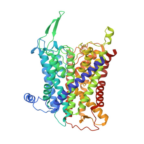
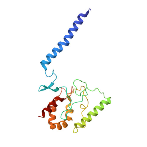
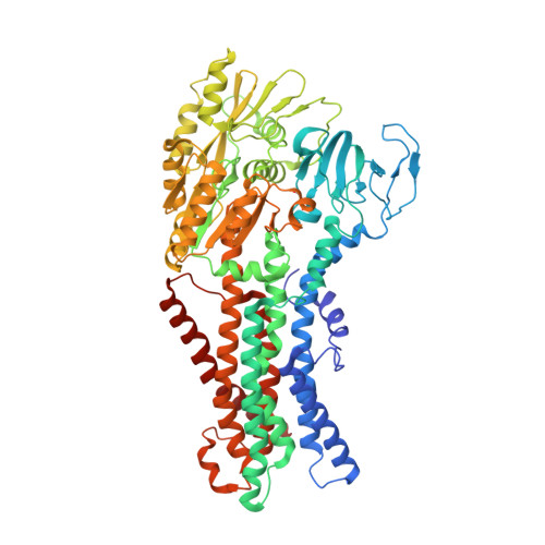
Cryo-EM map of the WT KdpFABC complex in the E1-P tight conformation, stabilised with the inhibitor orthovanadate


Experimental Data Snapshot

  • Method: ELECTRON MICROSCOPY
  • Resolution: 3.30 Å
  • Aggregation State: PARTICLE 
  • Reconstruction Method: SINGLE PARTICLE 

Starting Model: experimental
View more details

wwPDB Validation   3D Report Full Report


This is version 1.1 of the entry. See complete history


Literature

Inhibited KdpFABC transitions into an E1 off-cycle state.

Silberberg, J.M.Stock, C.Hielkema, L.Corey, R.A.Rheinberger, J.Wunnicke, D.Dubach, V.R.A.Stansfeld, P.J.Hanelt, I.Paulino, C.

(2022) Elife 11

  • DOI: https://doi.org/10.7554/eLife.80988
  • Primary Citation of Related Structures:  
    7ZRD, 7ZRE, 7ZRG, 7ZRH, 7ZRI, 7ZRJ, 7ZRK, 7ZRL, 7ZRM

  • PubMed Abstract: 

    KdpFABC is a high-affinity prokaryotic K + uptake system that forms a functional chimera between a channel-like subunit (KdpA) and a P-type ATPase (KdpB). At high K + levels, KdpFABC needs to be inhibited to prevent excessive K + accumulation to the point of toxicity. This is achieved by a phosphorylation of the serine residue in the TGES 162 motif in the A domain of the pump subunit KdpB (KdpB S162-P ). Here, we explore the structural basis of inhibition by KdpB S162 phosphorylation by determining the conformational landscape of KdpFABC under inhibiting and non-inhibiting conditions. Under turnover conditions, we identified a new inhibited KdpFABC state that we termed E1P tight, which is not part of the canonical Post-Albers transport cycle of P-type ATPases. It likely represents the biochemically described stalled E1P state adopted by KdpFABC upon KdpB S162 phosphorylation. The E1P tight state exhibits a compact fold of the three cytoplasmic domains and is likely adopted when the transition from high-energy E1P states to E2P states is unsuccessful. This study represents a structural characterization of a biologically relevant off-cycle state in the P-type ATPase family and supports the emerging discussion of P-type ATPase regulation by such states.


  • Organizational Affiliation

    Institute of Biochemistry, Biocenter, Goethe University Frankfurt, Frankfurt, Germany.


Macromolecules
Find similar proteins by:  (by identity cutoff)  |  3D Structure
Entity ID: 1
MoleculeChains Sequence LengthOrganismDetailsImage
Potassium-transporting ATPase potassium-binding subunit557Escherichia coliMutation(s): 0 
Gene Names: kdpAb0698JW0686
UniProt
Find proteins for P03959 (Escherichia coli (strain K12))
Go to UniProtKB:  P03959
Sequence Annotations
Expand
  • Reference Sequence
Find similar proteins by:  (by identity cutoff)  |  3D Structure
Entity ID: 2
MoleculeChains Sequence LengthOrganismDetailsImage
Potassium-transporting ATPase KdpC subunitB [auth C]190Escherichia coliMutation(s): 0 
Gene Names: kdpCb0696JW0684
UniProt
Find proteins for P03961 (Escherichia coli (strain K12))
Go to UniProtKB:  P03961
Sequence Annotations
Expand
  • Reference Sequence
Find similar proteins by:  (by identity cutoff)  |  3D Structure
Entity ID: 3
MoleculeChains Sequence LengthOrganismDetailsImage
Potassium-transporting ATPase KdpF subunitC [auth D]29Escherichia coliMutation(s): 0 
Gene Names: kdpFb4513JW0687
UniProt
Find proteins for P36937 (Escherichia coli (strain K12))
Go to UniProtKB:  P36937
Sequence Annotations
Expand
  • Reference Sequence
Find similar proteins by:  (by identity cutoff)  |  3D Structure
Entity ID: 4
MoleculeChains Sequence LengthOrganismDetailsImage
Potassium-transporting ATPase ATP-binding subunitD [auth B]682Escherichia coliMutation(s): 0 
Gene Names: kdpBb0697JW0685
EC: 7.2.2.6
UniProt
Find proteins for P03960 (Escherichia coli (strain K12))
Go to UniProtKB:  P03960
Sequence Annotations
Expand
  • Reference Sequence
Small Molecules
Ligands 3 Unique
IDChains Name / Formula / InChI Key2D Diagram3D Interactions
CDL
Query on CDL

Download Ideal Coordinates CCD File 
L [auth A],
M [auth A]
CARDIOLIPIN
C81 H156 O17 P2
XVTUQDWPJJBEHJ-KZCWQMDCSA-L
VO4 (Subject of Investigation/LOI)
Query on VO4

Download Ideal Coordinates CCD File 
P [auth B]VANADATE ION
O4 V
LSGOVYNHVSXFFJ-UHFFFAOYSA-N
K
Query on K

Download Ideal Coordinates CCD File 
E [auth A]
F [auth A]
G [auth A]
H [auth A]
I [auth A]
E [auth A],
F [auth A],
G [auth A],
H [auth A],
I [auth A],
J [auth A],
K [auth A],
N [auth B],
O [auth B]
POTASSIUM ION
K
NPYPAHLBTDXSSS-UHFFFAOYSA-N
Modified Residues  1 Unique
IDChains TypeFormula2D DiagramParent
SEP
Query on SEP
D [auth B]L-PEPTIDE LINKINGC3 H8 N O6 PSER
Experimental Data & Validation

Experimental Data

  • Method: ELECTRON MICROSCOPY
  • Resolution: 3.30 Å
  • Aggregation State: PARTICLE 
  • Reconstruction Method: SINGLE PARTICLE 
EM Software:
TaskSoftware PackageVersion
RECONSTRUCTIONRELION3.0.8
MODEL REFINEMENTPHENIX

Structure Validation

View Full Validation Report


Entry History & Funding Information

Deposition Data


Funding OrganizationLocationGrant Number
Netherlands Organisation for Scientific Research (NWO)United Kingdom722.017.001
Netherlands Organisation for Scientific Research (NWO)United Kingdom740.018.016
German Research Foundation (DFG)United Kingdom6322/3
German Research Foundation (DFG)United KingdomCRC807
Wellcome TrustUnited Kingdom208361/Z/17/Z

Revision History  (Full details and data files)

  • Version 1.0: 2022-11-16
    Type: Initial release
  • Version 1.1: 2024-10-23
    Changes: Data collection, Refinement description, Structure summary