7ZPF | pdb_00007zpf

Three-dimensional structure of AIP56, a short-trip single chain AB toxin from Photobacterium damselae subsp. piscicida.


Experimental Data Snapshot

  • Method: X-RAY DIFFRACTION
  • Resolution: 2.54 Å
  • R-Value Free: 
    0.282 (Depositor), 0.288 (DCC) 
  • R-Value Work: 
    0.240 (Depositor), 0.236 (DCC) 
  • R-Value Observed: 
    0.242 (Depositor) 

Starting Model: in silico
View more details

wwPDB Validation 3D Report Full Report

Validation slider image for 7ZPF

This is version 1.5 of the entry. See complete history

Literature

Unconventional structure and mechanisms for membrane interaction and translocation of the NF-kappa B-targeting toxin AIP56.

Lisboa, J.Pereira, C.Pinto, R.D.Rodrigues, I.S.Pereira, L.M.G.Pinheiro, B.Oliveira, P.Pereira, P.J.B.Azevedo, J.E.Durand, D.Benz, R.do Vale, A.Dos Santos, N.M.S.

(2023) Nat Commun 14: 7431-7431

  • DOI: https://doi.org/10.1038/s41467-023-43054-z
  • Primary Citation Related Structures: 
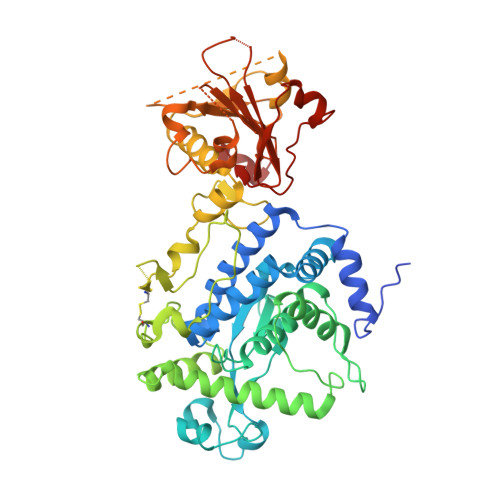
    7ZPF

  • PubMed Abstract: 

    Bacterial AB toxins are secreted key virulence factors that are internalized by target cells through receptor-mediated endocytosis, translocating their enzymatic domain to the cytosol from endosomes (short-trip) or the endoplasmic reticulum (long-trip). To accomplish this, bacterial AB toxins evolved a multidomain structure organized into either a single polypeptide chain or non-covalently associated polypeptide chains. The prototypical short-trip single-chain toxin is characterized by a receptor-binding domain that confers cellular specificity and a translocation domain responsible for pore formation whereby the catalytic domain translocates to the cytosol in an endosomal acidification-dependent way. In this work, the determination of the three-dimensional structure of AIP56 shows that, instead of a two-domain organization suggested by previous studies, AIP56 has three-domains: a non-LEE encoded effector C (NleC)-like catalytic domain associated with a small middle domain that contains the linker-peptide, followed by the receptor-binding domain. In contrast to prototypical single-chain AB toxins, AIP56 does not comprise a typical structurally complex translocation domain; instead, the elements involved in translocation are scattered across its domains. Thus, the catalytic domain contains a helical hairpin that serves as a molecular switch for triggering the conformational changes necessary for membrane insertion only upon endosomal acidification, whereas the middle and receptor-binding domains are required for pore formation.


  • Organizational Affiliation
    • Fish Immunology and Vaccinology Group, IBMC-Instituto de Biologia Molecular e Celular, Universidade do Porto, 4200-135, Porto, Portugal. johnny.lisboa@i3s.up.pt.

Macromolecule Content 

  • Total Structure Weight: 237 kDa 
  • Atom Count: 14,880 
  • Modeled Residue Count: 1,810 
  • Deposited Residue Count: 2,084 
  • Unique protein chains: 1

Macromolecules

Find similar proteins by:|  3D Structure
Entity ID: 1
MoleculeChains  Sequence LengthOrganismDetailsImage
Aip56
A, B, C, D
521Photobacterium damselae subsp. piscicidaMutation(s): 0 
Gene Names: aip56E4T25_16935
UniProt
Find proteins for Q2VL32 (Photobacterium damsela subsp. piscicida)
Explore Q2VL32 
Go to UniProtKB:  Q2VL32
Entity Groups
Sequence Clusters30% Identity50% Identity70% Identity90% Identity95% Identity100% Identity
UniProt GroupQ2VL32
Sequence Annotations
Expand
Reference Sequence

Small Molecules

Ligands 3 Unique
IDChains Name / Formula / InChI Key2D Diagram3D Interactions
GOL

Query on GOL


Download:Ideal Coordinates CCD File
G [auth A],
I [auth B],
J [auth B],
K [auth B],
L [auth B]
GLYCEROL
C3 H8 O3
PEDCQBHIVMGVHV-UHFFFAOYSA-N
ZN

Query on ZN


Download:Ideal Coordinates CCD File
E [auth A],
H [auth B],
M [auth C],
O [auth D]
ZINC ION
Zn
PTFCDOFLOPIGGS-UHFFFAOYSA-N
NI

Query on NI


Download:Ideal Coordinates CCD File
F [auth A],
N [auth C],
P [auth D],
Q [auth D]
NICKEL (II) ION
Ni
VEQPNABPJHWNSG-UHFFFAOYSA-N

Experimental Data & Validation

Experimental Data

  • Method: X-RAY DIFFRACTION
  • Resolution: 2.54 Å
  • R-Value Free:  0.282 (Depositor), 0.288 (DCC) 
  • R-Value Work:  0.240 (Depositor), 0.236 (DCC) 
  • R-Value Observed: 0.242 (Depositor) 
Space Group: P 1 21 1
Unit Cell:
Length ( Å )Angle ( ˚ )
a = 72.129α = 90
b = 193.85β = 113.24
c = 91.616γ = 90
Software Package:
Software NamePurpose
BUSTERrefinement
PHENIXrefinement
XDSdata reduction
STARANISOdata scaling
PHASERphasing

Structure Validation

View Full Validation Report


Entry History 

& Funding Information

Deposition Data


Funding OrganizationLocationGrant Number
Fundacao para a Ciencia e a TecnologiaPortugal268319 - PTDC/BIA-MIC/29910/2017-029910

Revision History  (Full details and data files)

  • Version 1.0: 2023-05-10
    Type: Initial release
  • Version 1.1: 2023-05-31
    Changes: Derived calculations
  • Version 1.2: 2023-06-28
    Changes: Derived calculations
  • Version 1.3: 2023-11-29
    Changes: Data collection, Database references
  • Version 1.4: 2024-05-01
    Changes: Refinement description
  • Version 1.5: 2024-11-13
    Changes: Structure summary