7ZOT | pdb_00007zot

crystal structure of PLAAT4 N-terminal domain


Experimental Data Snapshot

  • Method: X-RAY DIFFRACTION
  • Resolution: 1.74 Å
  • R-Value Free: 
    0.201 (Depositor), 0.201 (DCC) 
  • R-Value Work: 
    0.184 (Depositor), 0.184 (DCC) 

Starting Model: experimental
View more details

wwPDB Validation 3D Report Full Report

Validation slider image for 7ZOT

This is version 1.2 of the entry. See complete history

Literature

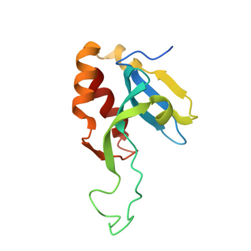
Crystal structure of the phospholipase A and acyltransferase 4 (PLAAT4) catalytic domain.

Wehlin, A.Cornaciu, I.Marquez, J.A.Perrakis, A.von Castelmur, E.

(2022) J Struct Biol 214: 107903-107903

  • DOI: https://doi.org/10.1016/j.jsb.2022.107903
  • Primary Citation Related Structures: 
    7ZOT

  • PubMed Abstract: 

    Phospholipase A and Acyltransferase 4 (PLAAT4) is a class II tumor suppressor, that also plays a role as a restrictor of intracellular Toxoplasma gondii infection through restriction of parasitic vacuole size. The catalytic N-terminal domain (NTD) interacts with the C-terminal domain (CTD), which is important for sub-cellular targeting and enzymatic function. The dynamics of the NTD main (L1) loop and the L2(B6) loop adjacent to the active site, have been shown to be important regulators of enzymatic activity. Here, we present the crystal structure of PLAAT4 NTD, determined from severely intergrown crystals using automated, laser-based crystal harvesting and data reduction technologies. The structure showed the L1 loop in two distinct conformations, highlighting a complex network of interactions likely influencing its conformational flexibility. Ensemble refinement of the crystal structure recapitulates the major correlated motions observed in solution by NMR. Our analysis offers useful insights on millisecond dynamics based on the crystal structure, complementing NMR studies which preclude structural information at this time scale.


  • Organizational Affiliation
    • Department of Physics, Chemistry and Biology, Linköping University, Sweden.

Macromolecule Content 

  • Total Structure Weight: 28.2 kDa 
  • Atom Count: 2,031 
  • Modeled Residue Count: 236 
  • Deposited Residue Count: 252 
  • Unique protein chains: 1

Macromolecules

Find similar proteins by:|  3D Structure
Entity ID: 1
MoleculeChains  Sequence LengthOrganismDetailsImage
Phospholipase A and acyltransferase 4
A, B
126Homo sapiensMutation(s): 0 
Gene Names: PLAAT4RARRES3RIG1TIG3
EC: 2.3.1 (PDB Primary Data), 3.1.1.32 (PDB Primary Data), 3.1.1.4 (PDB Primary Data)
UniProt & NIH Common Fund Data Resources
Find proteins for Q9UL19 (Homo sapiens)
Explore Q9UL19 
Go to UniProtKB:  Q9UL19
PHAROS:  Q9UL19
GTEx:  ENSG00000133321 
Entity Groups
Sequence Clusters30% Identity50% Identity70% Identity90% Identity95% Identity100% Identity
UniProt GroupQ9UL19
Sequence Annotations
Expand
Reference Sequence

Small Molecules

Ligands 1 Unique
IDChains Name / Formula / InChI Key2D Diagram3D Interactions
PEG

Query on PEG


Download:Ideal Coordinates CCD File
C [auth B]DI(HYDROXYETHYL)ETHER
C4 H10 O3
MTHSVFCYNBDYFN-UHFFFAOYSA-N

Experimental Data & Validation

Experimental Data

  • Method: X-RAY DIFFRACTION
  • Resolution: 1.74 Å
  • R-Value Free:  0.201 (Depositor), 0.201 (DCC) 
  • R-Value Work:  0.184 (Depositor), 0.184 (DCC) 
Space Group: C 1 2 1
Unit Cell:
Length ( Å )Angle ( ˚ )
a = 116.8α = 90
b = 39.54β = 94.952
c = 52.95γ = 90
Software Package:
Software NamePurpose
REFMACrefinement
autoPROCdata reduction
Aimlessdata scaling
PHASERphasing

Structure Validation

View Full Validation Report


Entry History 

& Funding Information

Deposition Data


Funding OrganizationLocationGrant Number
Swiss National Science FoundationSwitzerland--

Revision History  (Full details and data files)

  • Version 1.0: 2022-10-26
    Type: Initial release
  • Version 1.1: 2022-11-02
    Changes: Database references
  • Version 1.2: 2024-01-31
    Changes: Data collection, Refinement description