7ZKG | pdb_00007zkg

C-Methyltransferase PsmD from Streptomyces griseofuscus with bound cofactor (crystal form 2)


Experimental Data Snapshot

  • Method: X-RAY DIFFRACTION
  • Resolution: 2.30 Å
  • R-Value Free: 
    0.216 (Depositor), 0.187 (DCC) 
  • R-Value Work: 
    0.189 (Depositor) 
  • R-Value Observed: 
    0.190 (Depositor) 

Starting Model: experimental
View more details

wwPDB Validation 3D Report Full Report

Validation slider image for 7ZKG

Ligand Structure Quality Assessment 


This is version 1.2 of the entry. See complete history

Literature

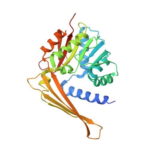
Enzymatic C3-Methylation of Indoles Using Methyltransferase PsmD-Crystal Structure, Catalytic Mechanism, and Preparative Applications

Amariei, D.A.Pozhydaieva, N.David, B.Schneider, P.Classen, T.Gohlke, H.Weiergraber, O.H.Pietruszka, J.

(2022) ACS Catal 

Macromolecule Content 

  • Total Structure Weight: 63.37 kDa 
  • Atom Count: 4,163 
  • Modeled Residue Count: 504 
  • Deposited Residue Count: 550 
  • Unique protein chains: 1

Macromolecules

Find similar proteins by:|  3D Structure
Entity ID: 1
MoleculeChains  Sequence LengthOrganismDetailsImage
Methyltransferase
A, B
275Streptomyces griseofuscusMutation(s): 0 
Gene Names: psmD
UniProt
Find proteins for W8R3D8 (Streptomyces griseofuscus)
Explore W8R3D8 
Go to UniProtKB:  W8R3D8
Entity Groups
Sequence Clusters30% Identity50% Identity70% Identity90% Identity95% Identity100% Identity
UniProt GroupW8R3D8
Sequence Annotations
Expand
Reference Sequence

Small Molecules

Ligands 3 Unique
IDChains Name / Formula / InChI Key2D Diagram3D Interactions
SAH
(Subject of Investigation/LOI)

Query on SAH


Download:Ideal Coordinates CCD File
J [auth A],
N [auth B]
S-ADENOSYL-L-HOMOCYSTEINE
C14 H20 N6 O5 S
ZJUKTBDSGOFHSH-WFMPWKQPSA-N
MPD

Query on MPD


Download:Ideal Coordinates CCD File
H [auth A],
I [auth A],
L [auth B],
M [auth B]
(4S)-2-METHYL-2,4-PENTANEDIOL
C6 H14 O2
SVTBMSDMJJWYQN-YFKPBYRVSA-N
PO4

Query on PO4


Download:Ideal Coordinates CCD File
C [auth A]
D [auth A]
E [auth A]
F [auth A]
G [auth A]
C [auth A],
D [auth A],
E [auth A],
F [auth A],
G [auth A],
K [auth B]
PHOSPHATE ION
O4 P
NBIIXXVUZAFLBC-UHFFFAOYSA-K

Experimental Data & Validation

Experimental Data

  • Method: X-RAY DIFFRACTION
  • Resolution: 2.30 Å
  • R-Value Free:  0.216 (Depositor), 0.187 (DCC) 
  • R-Value Work:  0.189 (Depositor) 
  • R-Value Observed: 0.190 (Depositor) 
Space Group: P 61 2 2
Unit Cell:
Length ( Å )Angle ( ˚ )
a = 138.71α = 90
b = 138.71β = 90
c = 162.06γ = 120
Software Package:
Software NamePurpose
PHENIXrefinement
XDSdata reduction
XDSdata scaling
MOLREPphasing

Structure Validation

View Full Validation Report


Ligand Structure Quality Assessment 


Entry History 

& Funding Information

Deposition Data


Funding OrganizationLocationGrant Number
Not funded--

Revision History  (Full details and data files)

  • Version 1.0: 2022-12-28
    Type: Initial release
  • Version 1.1: 2024-01-31
    Changes: Data collection, Refinement description
  • Version 1.2: 2025-12-10
    Changes: Data collection, Structure summary