7ZI0 | pdb_00007zi0

Structure of human Smoothened in complex with cholesterol and SAG


Experimental Data Snapshot

  • Method: X-RAY DIFFRACTION
  • Resolution: 3.00 Å
  • R-Value Free: 
    0.275 (Depositor), 0.270 (DCC) 
  • R-Value Work: 
    0.231 (Depositor), 0.232 (DCC) 
  • R-Value Observed: 
    0.233 (Depositor) 

Starting Model: experimental
View more details

wwPDB Validation 3D Report Full Report

Validation slider image for 7ZI0

Ligand Structure Quality Assessment 


This is version 1.2 of the entry. See complete history

Literature

Patched 1 regulates Smoothened by controlling sterol binding to its extracellular cysteine-rich domain.

Kinnebrew, M.Woolley, R.E.Ansell, T.B.Byrne, E.F.X.Frigui, S.Luchetti, G.Sircar, R.Nachtergaele, S.Mydock-McGrane, L.Krishnan, K.Newstead, S.Sansom, M.S.P.Covey, D.F.Siebold, C.Rohatgi, R.

(2022) Sci Adv 8: eabm5563-eabm5563

  • DOI: https://doi.org/10.1126/sciadv.abm5563
  • Primary Citation Related Structures: 
    7ZI0

  • PubMed Abstract: 

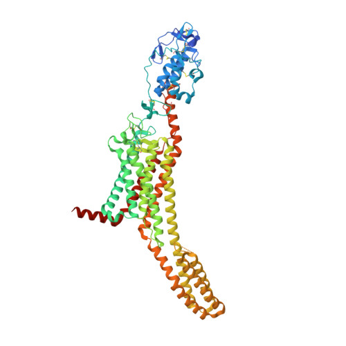
    Smoothened (SMO) transduces the Hedgehog (Hh) signal across the plasma membrane in response to accessible cholesterol. Cholesterol binds SMO at two sites: one in the extracellular cysteine-rich domain (CRD) and a second in the transmembrane domain (TMD). How these two sterol-binding sites mediate SMO activation in response to the ligand Sonic Hedgehog (SHH) remains unknown. We find that mutations in the CRD (but not the TMD) reduce the fold increase in SMO activity triggered by SHH. SHH also promotes the photocrosslinking of a sterol analog to the CRD in intact cells. In contrast, sterol binding to the TMD site boosts SMO activity regardless of SHH exposure. Mutational and computational analyses show that these sites are in allosteric communication despite being 45 angstroms apart. Hence, sterols function as both SHH-regulated orthosteric ligands at the CRD and allosteric ligands at the TMD to regulate SMO activity and Hh signaling.


  • Organizational Affiliation
    • Departments of Biochemistry and Medicine, Stanford University School of Medicine, Stanford, CA, USA.

Macromolecule Content 

  • Total Structure Weight: 143.95 kDa 
  • Atom Count: 9,409 
  • Modeled Residue Count: 1,171 
  • Deposited Residue Count: 1,276 
  • Unique protein chains: 1

Macromolecules

Find similar proteins by:|  3D Structure
Entity ID: 1
MoleculeChains  Sequence LengthOrganismDetailsImage
Smoothened homolog,Soluble cytochrome b562
A, B
638Homo sapiensMutation(s): 0 
Gene Names: SMOSMOHcybC
Membrane Entity: Yes 
UniProt & NIH Common Fund Data Resources
Find proteins for P0ABE7 (Escherichia coli)
Explore P0ABE7 
Go to UniProtKB:  P0ABE7
Find proteins for Q99835 (Homo sapiens)
Explore Q99835 
Go to UniProtKB:  Q99835
PHAROS:  Q99835
GTEx:  ENSG00000128602 
Entity Groups
Sequence Clusters30% Identity50% Identity70% Identity90% Identity95% Identity100% Identity
UniProt GroupsP0ABE7Q99835
Glycosylation
Glycosylation Sites: 1Go to GlyGen: Q99835-1
Sequence Annotations
Expand
Reference Sequence

Small Molecules

Ligands 5 Unique
IDChains Name / Formula / InChI Key2D Diagram3D Interactions
V0S
(Subject of Investigation/LOI)

Query on V0S


Download:Ideal Coordinates CCD File
C [auth A],
I [auth B]
3-chloro-N-[trans-4-(methylamino)cyclohexyl]-N-{[3-(pyridin-4-yl)phenyl]methyl}-1-benzothiophene-2-carboxamide
C28 H28 Cl N3 O S
VFSUUTYAEQOIMW-YHBQERECSA-N
CLR
(Subject of Investigation/LOI)

Query on CLR


Download:Ideal Coordinates CCD File
D [auth A],
J [auth B]
CHOLESTEROL
C27 H46 O
HVYWMOMLDIMFJA-DPAQBDIFSA-N
MPG
(Subject of Investigation/LOI)

Query on MPG


Download:Ideal Coordinates CCD File
H [auth A][(Z)-octadec-9-enyl] (2R)-2,3-bis(oxidanyl)propanoate
C21 H40 O4
JPJYKWFFJCWMPK-GDCKJWNLSA-N
NAG
(Subject of Investigation/LOI)

Query on NAG


Download:Ideal Coordinates CCD File
E [auth A]2-acetamido-2-deoxy-beta-D-glucopyranose
C8 H15 N O6
OVRNDRQMDRJTHS-FMDGEEDCSA-N
NA
(Subject of Investigation/LOI)

Query on NA


Download:Ideal Coordinates CCD File
F [auth A],
G [auth A]
SODIUM ION
Na
FKNQFGJONOIPTF-UHFFFAOYSA-N

Experimental Data & Validation

Experimental Data

  • Method: X-RAY DIFFRACTION
  • Resolution: 3.00 Å
  • R-Value Free:  0.275 (Depositor), 0.270 (DCC) 
  • R-Value Work:  0.231 (Depositor), 0.232 (DCC) 
  • R-Value Observed: 0.233 (Depositor) 
Space Group: C 1 2 1
Unit Cell:
Length ( Å )Angle ( ˚ )
a = 122.86α = 90
b = 63.15β = 96.29
c = 208.06γ = 90
Software Package:
Software NamePurpose
REFMACrefinement
PDB_EXTRACTdata extraction
DIALSdata reduction
Aimlessdata scaling
PHASERphasing

Structure Validation

View Full Validation Report


Ligand Structure Quality Assessment 


Entry History 

& Funding Information

Deposition Data


Funding OrganizationLocationGrant Number
Cancer Research UKUnited KingdomC20724/A26752
European Research Council (ERC)European Union647278

Revision History  (Full details and data files)

  • Version 1.0: 2022-06-15
    Type: Initial release
  • Version 1.1: 2024-01-31
    Changes: Data collection, Refinement description
  • Version 1.2: 2024-10-23
    Changes: Structure summary