7ZHT | pdb_00007zht

Leishmania donovani Glucose 6-Phosphate Dehydrogenase apo form


Experimental Data Snapshot

  • Method: X-RAY DIFFRACTION
  • Resolution: 2.80 Å
  • R-Value Free: 
    0.304 (Depositor), 0.302 (DCC) 
  • R-Value Work: 
    0.244 (Depositor), 0.244 (DCC) 
  • R-Value Observed: 
    0.250 (Depositor) 

Starting Model: experimental
View more details

wwPDB Validation 3D Report Full Report

Validation slider image for 7ZHT

This is version 1.3 of the entry. See complete history

Literature

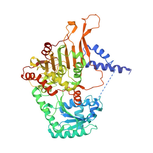
Crystal structure of Leishmania donovani glucose 6-phosphate dehydrogenase reveals a unique N-terminal domain.

Berneburg, I.Rahlfs, S.Becker, K.Fritz-Wolf, K.

(2022) Commun Biol 5: 1353-1353

  • DOI: https://doi.org/10.1038/s42003-022-04307-7
  • Primary Citation Related Structures: 
    7ZHT, 7ZHU, 7ZHV, 7ZHW, 7ZHX, 7ZHY, 7ZHZ

  • PubMed Abstract: 

    Since unicellular parasites highly depend on NADPH as a source for reducing equivalents, the pentose phosphate pathway, especially the first and rate-limiting NADPH-producing enzyme glucose 6-phosphate dehydrogenase (G6PD), is considered an excellent antitrypanosomatid drug target. Here we present the crystal structure of Leishmania donovani G6PD (LdG6PD) elucidating the unique N-terminal domain of Kinetoplastida G6PDs. Our investigations on the function of the N-domain suggest its involvement in the formation of a tetramer that is completely different from related Trypanosoma G6PDs. Structural and functional investigations further provide interesting insights into the binding mode of LdG6PD, following an ordered mechanism, which is confirmed by a G6P-induced domain shift and rotation of the helical N-domain. Taken together, these insights into LdG6PD contribute to the understanding of G6PDs' molecular mechanisms and provide an excellent basis for further drug discovery approaches.


  • Organizational Affiliation
    • Biochemistry and Molecular Biology, Interdisciplinary Research Center, Justus Liebig University, Giessen, Germany.

Macromolecule Content 

  • Total Structure Weight: 254.48 kDa 
  • Atom Count: 16,104 
  • Modeled Residue Count: 2,023 
  • Deposited Residue Count: 2,248 
  • Unique protein chains: 1

Macromolecules

Find similar proteins by:|  3D Structure
Entity ID: 1
MoleculeChains  Sequence LengthOrganismDetailsImage
Glucose-6-phosphate 1-dehydrogenase
A, B, C, D
562Leishmania donovaniMutation(s): 0 
Gene Names: g6pdh
EC: 1.1.1.49
UniProt
Find proteins for A2CIL3 (Leishmania donovani)
Explore A2CIL3 
Go to UniProtKB:  A2CIL3
Entity Groups
Sequence Clusters30% Identity50% Identity70% Identity90% Identity95% Identity100% Identity
UniProt GroupA2CIL3
Sequence Annotations
Expand
Reference Sequence

Small Molecules

Ligands 2 Unique
IDChains Name / Formula / InChI Key2D Diagram3D Interactions
SO4

Query on SO4


Download:Ideal Coordinates CCD File
G [auth A],
K [auth B],
O [auth C]
SULFATE ION
O4 S
QAOWNCQODCNURD-UHFFFAOYSA-L
EDO

Query on EDO


Download:Ideal Coordinates CCD File
E [auth A]
F [auth A]
H [auth B]
I [auth B]
J [auth B]
E [auth A],
F [auth A],
H [auth B],
I [auth B],
J [auth B],
L [auth C],
M [auth C],
N [auth C],
P [auth D]
1,2-ETHANEDIOL
C2 H6 O2
LYCAIKOWRPUZTN-UHFFFAOYSA-N

Experimental Data & Validation

Experimental Data

  • Method: X-RAY DIFFRACTION
  • Resolution: 2.80 Å
  • R-Value Free:  0.304 (Depositor), 0.302 (DCC) 
  • R-Value Work:  0.244 (Depositor), 0.244 (DCC) 
  • R-Value Observed: 0.250 (Depositor) 
Space Group: P 1 2 1
Unit Cell:
Length ( Å )Angle ( ˚ )
a = 117.69α = 90
b = 65.82β = 92.35
c = 189.22γ = 90
Software Package:
Software NamePurpose
PHENIXrefinement
PHENIXrefinement
XDSdata reduction
XSCALEdata scaling
PHENIXphasing

Structure Validation

View Full Validation Report


Entry History 

& Funding Information

Deposition Data


Funding OrganizationLocationGrant Number
LOEWE Center DRUID (Project B3 and E3) within the Hessian Excellence ProgramGermanyProject B3 and E3

Revision History  (Full details and data files)

  • Version 1.0: 2022-12-14
    Type: Initial release
  • Version 1.1: 2022-12-21
    Changes: Database references
  • Version 1.2: 2024-01-31
    Changes: Data collection, Refinement description
  • Version 1.3: 2025-10-01
    Changes: Advisory, Derived calculations, Structure summary