7ZFR | pdb_00007zfr

Crystal structure of HLA-DP (DPA1*02:01-DPB1*01:01) in complex with a peptide bound in the reverse direction


Experimental Data Snapshot

  • Method: X-RAY DIFFRACTION
  • Resolution: 2.90 Å
  • R-Value Free: 
    0.272 (Depositor), 0.272 (DCC) 
  • R-Value Work: 
    0.231 (Depositor), 0.231 (DCC) 
  • R-Value Observed: 
    0.235 (Depositor) 

Starting Model: experimental
View more details

wwPDB Validation 3D Report Full Report

Validation slider image for 7ZFR

This is version 1.4 of the entry. See complete history

Literature

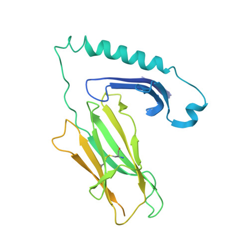
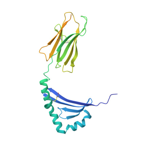
Machine learning predictions of MHC-II specificities reveal alternative binding mode of class II epitopes.

Racle, J.Guillaume, P.Schmidt, J.Michaux, J.Larabi, A.Lau, K.Perez, M.A.S.Croce, G.Genolet, R.Coukos, G.Zoete, V.Pojer, F.Bassani-Sternberg, M.Harari, A.Gfeller, D.

(2023) Immunity 56: 1359-1375.e13

  • DOI: https://doi.org/10.1016/j.immuni.2023.03.009
  • Primary Citation Related Structures: 
    7ZAK, 7ZFR

  • PubMed Abstract: 

    CD4 + T cells orchestrate the adaptive immune response against pathogens and cancer by recognizing epitopes presented on class II major histocompatibility complex (MHC-II) molecules. The high polymorphism of MHC-II genes represents an important hurdle toward accurate prediction and identification of CD4 + T cell epitopes. Here we collected and curated a dataset of 627,013 unique MHC-II ligands identified by mass spectrometry. This enabled us to precisely determine the binding motifs of 88 MHC-II alleles across humans, mice, cattle, and chickens. Analysis of these binding specificities combined with X-ray crystallography refined our understanding of the molecular determinants of MHC-II motifs and revealed a widespread reverse-binding mode in HLA-DP ligands. We then developed a machine-learning framework to accurately predict binding specificities and ligands of any MHC-II allele. This tool improves and expands predictions of CD4 + T cell epitopes and enables us to discover viral and bacterial epitopes following the aforementioned reverse-binding mode.


  • Organizational Affiliation
    • Department of Oncology UNIL CHUV, Ludwig Institute for Cancer Research, University of Lausanne, Lausanne, Switzerland; Swiss Institute of Bioinformatics (SIB), Lausanne, Switzerland; Agora Cancer Research Centre, Lausanne, Switzerland; Swiss Cancer Center Leman (SCCL), Lausanne, Switzerland. Electronic address: julien.racle@unil.ch.

Macromolecule Content 

  • Total Structure Weight: 61.44 kDa 
  • Atom Count: 3,156 
  • Modeled Residue Count: 381 
  • Deposited Residue Count: 542 
  • Unique protein chains: 3

Macromolecules

Find similar proteins by:|  3D Structure
Entity ID: 1
MoleculeChains  Sequence LengthOrganismDetailsImage
MHC class II HLA-DP alpha chain (DPA1*02:01)268Homo sapiensMutation(s): 0 
Gene Names: HLA-DPA1
Find similar proteins by:|  3D Structure
Entity ID: 2
MoleculeChains  Sequence LengthOrganismDetailsImage
MHC class II HLA-DP beta chain (DPB1*01:01)262Homo sapiensMutation(s): 0 
Gene Names: HLA-DPB1
Find similar proteins by:  Sequence   |   3D Structure  
Entity ID: 3
MoleculeChains  Sequence LengthOrganismDetailsImage
Synthetic peptide12Homo sapiensMutation(s): 0 

Oligosaccharides

Help  
Entity ID: 4
MoleculeChains Length2D Diagram GlycosylationD Interactions
2-acetamido-2-deoxy-beta-D-glucopyranose-(1-4)-2-acetamido-2-deoxy-beta-D-glucopyranose
D
2N-Glycosylation
Glycosylation Resources
GlyTouCan: G42666HT
GlyCosmos: G42666HT
GlyGen: G42666HT

Experimental Data & Validation

Experimental Data

  • Method: X-RAY DIFFRACTION
  • Resolution: 2.90 Å
  • R-Value Free:  0.272 (Depositor), 0.272 (DCC) 
  • R-Value Work:  0.231 (Depositor), 0.231 (DCC) 
  • R-Value Observed: 0.235 (Depositor) 
Space Group: P 61 2 2
Unit Cell:
Length ( Å )Angle ( ˚ )
a = 83.985α = 90
b = 83.985β = 90
c = 348.589γ = 120
Software Package:
Software NamePurpose
PHENIXrefinement
XDSdata reduction
XDSdata scaling
PHASERphasing

Structure Validation

View Full Validation Report


Entry History 

& Funding Information

Deposition Data


Funding OrganizationLocationGrant Number
Swiss Cancer LeagueSwitzerlandKFS-4961-02-2020

Revision History  (Full details and data files)

  • Version 1.0: 2023-04-12
    Type: Initial release
  • Version 1.1: 2023-04-19
    Changes: Database references
  • Version 1.2: 2023-06-21
    Changes: Database references
  • Version 1.3: 2024-02-07
    Changes: Data collection, Refinement description
  • Version 1.4: 2024-10-23
    Changes: Structure summary