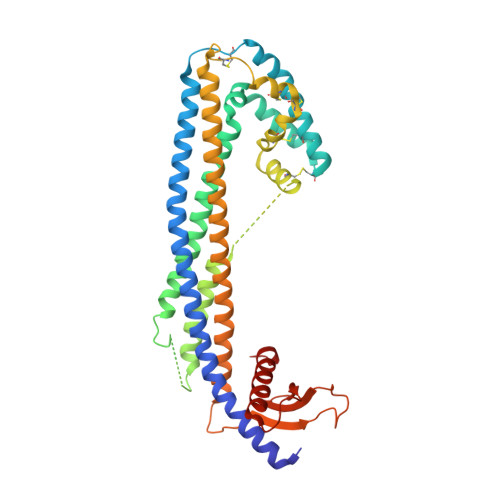
Hydrophobic tails enable diverse functions of the extracellular chaperone clusterin
Yuste-Checa, P., Carvajal, A.I., Mi, C., Paatz, S., Ulrich Hartl, F., Bracher, A.(2024) bioRxiv
Experimental Data Snapshot
(2024) bioRxiv
Entity ID: 1 | |||||
|---|---|---|---|---|---|
| Molecule | Chains | Sequence Length | Organism | Details | Image |
| Clusterin | A, B [auth D] | 402 | Homo sapiens | Mutation(s): 0 Gene Names: CLU, APOJ, CLI, KUB1, AAG4 |  |
UniProt & NIH Common Fund Data Resources | |||||
Find proteins for P10909 (Homo sapiens) Explore P10909 Go to UniProtKB: P10909 | |||||
PHAROS: P10909 GTEx: ENSG00000120885 | |||||
Entity Groups | |||||
| Sequence Clusters | 30% Identity50% Identity70% Identity90% Identity95% Identity100% Identity | ||||
| UniProt Group | P10909 | ||||
Glycosylation | |||||
| Glycosylation Sites: 6 | Go to GlyGen: P10909-1 | ||||
Sequence AnnotationsExpand | |||||
| |||||
| Ligands 1 Unique | |||||
|---|---|---|---|---|---|
| ID | Chains | Name / Formula / InChI Key | 2D Diagram | 3D Interactions | |
| NAG Query on NAG | H [auth A] I [auth A] J [auth A] K [auth A] L [auth A] | 2-acetamido-2-deoxy-beta-D-glucopyranose C8 H15 N O6 OVRNDRQMDRJTHS-FMDGEEDCSA-N |  | ||
| Length ( Å ) | Angle ( ˚ ) |
|---|---|
| a = 194.44 | α = 90 |
| b = 46.438 | β = 127.2 |
| c = 155.173 | γ = 90 |
| Software Name | Purpose |
|---|---|
| XDS | data reduction |
| Aimless | data scaling |
| MOLREP | phasing |
| PHENIX | refinement |
| PDB_EXTRACT | data extraction |
| Funding Organization | Location | Grant Number |
|---|---|---|
| Not funded | -- |