7ZDK

IF(apo/asym) conformation of CydDC in AMP-PNP(CydC)/AMP-PNP(CydD) bound state (Dataset-8)


Experimental Data Snapshot

  • Method: ELECTRON MICROSCOPY
  • Resolution: 3.01 Å
  • Aggregation State: PARTICLE 
  • Reconstruction Method: SINGLE PARTICLE 

wwPDB Validation   3D Report Full Report


This is version 1.1 of the entry. See complete history


Literature

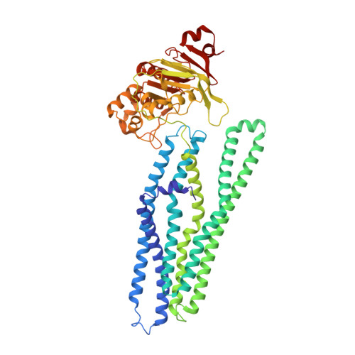
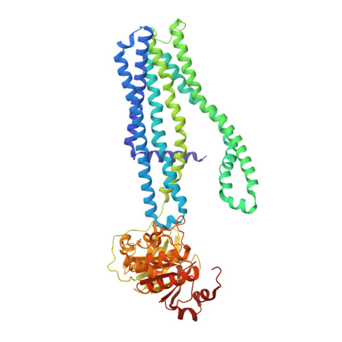
Dissecting the conformational complexity and mechanism of a bacterial heme transporter.

Wu, D.Mehdipour, A.R.Finke, F.Goojani, H.G.Groh, R.R.Grund, T.N.Reichhart, T.M.B.Zimmermann, R.Welsch, S.Bald, D.Shepherd, M.Hummer, G.Safarian, S.

(2023) Nat Chem Biol 19: 992-1003

  • DOI: https://doi.org/10.1038/s41589-023-01314-5
  • Primary Citation of Related Structures:  
    7ZD5, 7ZDA, 7ZDB, 7ZDC, 7ZDE, 7ZDF, 7ZDG, 7ZDK, 7ZDL, 7ZDR, 7ZDS, 7ZDT, 7ZDU, 7ZDV, 7ZDW, 7ZE5, 7ZEC

  • PubMed Abstract: 

    Iron-bound cyclic tetrapyrroles (hemes) are redox-active cofactors in bioenergetic enzymes. However, the mechanisms of heme transport and insertion into respiratory chain complexes remain unclear. Here, we used cellular, biochemical, structural and computational methods to characterize the structure and function of the heterodimeric bacterial ABC transporter CydDC. We provide multi-level evidence that CydDC is a heme transporter required for functional maturation of cytochrome bd, a pharmaceutically relevant drug target. Our systematic single-particle cryogenic-electron microscopy approach combined with atomistic molecular dynamics simulations provides detailed insight into the conformational landscape of CydDC during substrate binding and occlusion. Our simulations reveal that heme binds laterally from the membrane space to the transmembrane region of CydDC, enabled by a highly asymmetrical inward-facing CydDC conformation. During the binding process, heme propionates interact with positively charged residues on the surface and later in the substrate-binding pocket of the transporter, causing the heme orientation to rotate 180°.


  • Organizational Affiliation

    Department of Molecular Membrane Biology, Max Planck Institute of Biophysics, Frankfurt/Main, Germany.


Macromolecules
Find similar proteins by:  (by identity cutoff)  |  3D Structure
Entity ID: 1
MoleculeChains Sequence LengthOrganismDetailsImage
ATP-binding/permease protein CydCA [auth C]573Escherichia coli K-12Mutation(s): 0 
Gene Names: cydCmdrAmdrHsurBb0886JW0869
Membrane Entity: Yes 
UniProt
Find proteins for P23886 (Escherichia coli (strain K12))
Explore P23886 
Go to UniProtKB:  P23886
Entity Groups  
Sequence Clusters30% Identity50% Identity70% Identity90% Identity95% Identity100% Identity
UniProt GroupP23886
Sequence Annotations
Expand
  • Reference Sequence
Find similar proteins by:  (by identity cutoff)  |  3D Structure
Entity ID: 2
MoleculeChains Sequence LengthOrganismDetailsImage
ATP-binding/permease protein CydDB [auth D]588Escherichia coli K-12Mutation(s): 0 
Gene Names: cydDhtrDb0887JW0870
Membrane Entity: Yes 
UniProt
Find proteins for P29018 (Escherichia coli (strain K12))
Explore P29018 
Go to UniProtKB:  P29018
Entity Groups  
Sequence Clusters30% Identity50% Identity70% Identity90% Identity95% Identity100% Identity
UniProt GroupP29018
Sequence Annotations
Expand
  • Reference Sequence
Experimental Data & Validation

Experimental Data

  • Method: ELECTRON MICROSCOPY
  • Resolution: 3.01 Å
  • Aggregation State: PARTICLE 
  • Reconstruction Method: SINGLE PARTICLE 

Structure Validation

View Full Validation Report


Entry History & Funding Information

Deposition Data


Funding OrganizationLocationGrant Number
Max Planck SocietyGermany--

Revision History  (Full details and data files)

  • Version 1.0: 2023-04-19
    Type: Initial release
  • Version 1.1: 2023-11-01
    Changes: Data collection, Database references