7ZCV | pdb_00007zcv

Rgg144 of Streptococcus pneumoniae


Experimental Data Snapshot

  • Method: X-RAY DIFFRACTION
  • Resolution: 1.90 Å
  • R-Value Free: 
    0.215 (Depositor), 0.211 (DCC) 
  • R-Value Work: 
    0.189 (Depositor) 
  • R-Value Observed: 
    0.190 (Depositor) 

wwPDB Validation 3D Report Full Report

Validation slider image for 7ZCV

This is version 1.3 of the entry. See complete history

Literature

Structure-function analysis for the development of peptide inhibitors for a Gram-positive quorum sensing system.

Abdullah, I.T.Ulijasz, A.T.Girija, U.V.Tam, S.Andrew, P.Hiller, N.L.Wallis, R.Yesilkaya, H.

(2022) Mol Microbiol 117: 1464-1478

  • DOI: https://doi.org/10.1111/mmi.14921
  • Primary Citation Related Structures: 
    7ZCV

  • PubMed Abstract: 

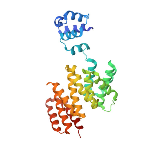
    The Streptococcus pneumoniae Rgg144/SHP144 regulator-peptide quorum sensing (QS) system is critical for nutrient utilization, oxidative stress response, and virulence. Here, we characterized this system by assessing the importance of each residue within the active short hydrophobic peptide (SHP) by alanine-scanning mutagenesis and testing the resulting peptides for receptor binding and activation of the receptor. Interestingly, several of the mutations had little effect on binding to Rgg144 but reduced transcriptional activation appreciably. In particular, a proline substitution (P21A) reduced transcriptional activation by 29-fold but bound with a 3-fold higher affinity than the wild-type SHP. Consistent with the function of Rgg144, the mutant peptide led to decreased utilization of mannose and increased susceptibility to superoxide generator paraquat. Pangenome comparison showed full conservation of P21 across SHP144 allelic variants. Crystallization of Rgg144 in the absence of peptide revealed a comparable structure to the DNA bound and free forms of its homologs suggesting similar mechanisms of activation. Together, these analyses identify key interactions in a critical pneumococcal QS system. Further manipulation of the SHP has the potential to facilitate the development of inhibitors that are functional across strains. The approach described here is likely to be effective across QS systems in multiple species.


  • Organizational Affiliation
    • Department of Respiratory Sciences, University of Leicester, Leicester, UK.

Macromolecule Content 

  • Total Structure Weight: 67.97 kDa 
  • Atom Count: 5,142 
  • Modeled Residue Count: 568 
  • Deposited Residue Count: 574 
  • Unique protein chains: 1

Macromolecules

Find similar proteins by:|  3D Structure
Entity ID: 1
MoleculeChains  Sequence LengthOrganismDetailsImage
Transcriptional regulator
A, B
287Streptococcus pneumoniaeMutation(s): 0 
Gene Names: mutRERS021218_00146ERS096071_01044SAMEA3381574_00832SAMEA3389353_01155
UniProt
Find proteins for A0A0E7KI02 (Streptococcus pneumoniae)
Explore A0A0E7KI02 
Go to UniProtKB:  A0A0E7KI02
Entity Groups
Sequence Clusters30% Identity50% Identity70% Identity90% Identity95% Identity100% Identity
UniProt GroupA0A0E7KI02
Sequence Annotations
Expand
Reference Sequence

Experimental Data & Validation

Experimental Data

  • Method: X-RAY DIFFRACTION
  • Resolution: 1.90 Å
  • R-Value Free:  0.215 (Depositor), 0.211 (DCC) 
  • R-Value Work:  0.189 (Depositor) 
  • R-Value Observed: 0.190 (Depositor) 
Space Group: P 21 21 21
Unit Cell:
Length ( Å )Angle ( ˚ )
a = 75.479α = 90
b = 80.871β = 90
c = 106.534γ = 90
Software Package:
Software NamePurpose
XDSdata reduction
PHENIXrefinement
PDB_EXTRACTdata extraction
xia2data scaling
PHASERphasing

Structure Validation

View Full Validation Report


Entry History 

& Funding Information

Deposition Data


Funding OrganizationLocationGrant Number
Not fundedUnited Kingdom--

Revision History  (Full details and data files)

  • Version 1.0: 2022-06-01
    Type: Initial release
  • Version 1.1: 2022-06-15
    Changes: Database references
  • Version 1.2: 2022-07-06
    Changes: Database references
  • Version 1.3: 2024-06-19
    Changes: Data collection, Refinement description