


Serial synchrotron crystallography structure of the Sensory rhodopsin II transducer complex
Ortolani, G., Bosman, R., Ostojic, L., Branden, G., Neutze, R.To be published.
Experimental Data Snapshot
Starting Model: experimental
View more details
Entity ID: 1 | |||||
|---|---|---|---|---|---|
| Molecule | Chains | Sequence Length | Organism | Details | Image |
| Sensory rhodopsin-2 | 225 | Natronomonas pharaonis | Mutation(s): 0 Gene Names: sop2, sopII Membrane Entity: Yes | 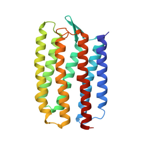 | |
UniProt | |||||
Entity Groups | |||||
| Sequence Clusters | 30% Identity50% Identity70% Identity90% Identity95% Identity100% Identity | ||||
| UniProt Group | P42196 | ||||
Sequence AnnotationsExpand | |||||
Reference Sequence | |||||
Entity ID: 2 | |||||
|---|---|---|---|---|---|
| Molecule | Chains | Sequence Length | Organism | Details | Image |
| Sensory rhodopsin II transducer | 66 | Natronomonas pharaonis | Mutation(s): 0 Gene Names: htr2, htrII Membrane Entity: Yes | 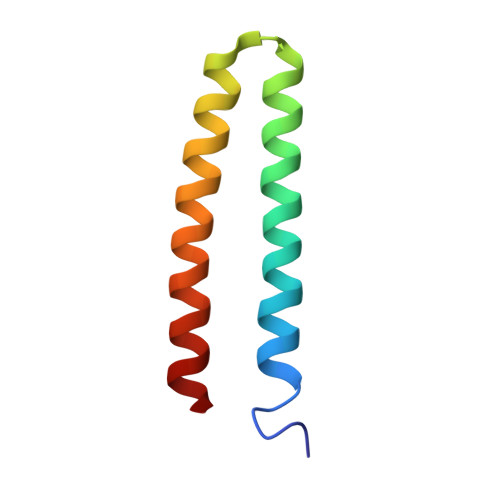 | |
UniProt | |||||
Entity Groups | |||||
| Sequence Clusters | 30% Identity50% Identity70% Identity90% Identity95% Identity100% Identity | ||||
| UniProt Group | P42259 | ||||
Sequence AnnotationsExpand | |||||
Reference Sequence | |||||
| Ligands 1 Unique | |||||
|---|---|---|---|---|---|
| ID | Chains | Name / Formula / InChI Key | 2D Diagram | 3D Interactions | |
| RET (Subject of Investigation/LOI) Download:Ideal Coordinates CCD File | C [auth A] | RETINAL C20 H28 O NCYCYZXNIZJOKI-OVSJKPMPSA-N |  | ||
| Length ( Å ) | Angle ( ˚ ) |
|---|---|
| a = 52.87 | α = 90 |
| b = 67.65 | β = 90 |
| c = 114.42 | γ = 90 |
| Software Name | Purpose |
|---|---|
| PHENIX | refinement |
| PDB_EXTRACT | data extraction |
| PHASER | phasing |
| careless | data reduction |
| careless | data scaling |
| Funding Organization | Location | Grant Number |
|---|---|---|
| H2020 Marie Curie Actions of the European Commission | European Union | X-Probe |
| Swedish Research Council | Sweden | 2015-00560 |


