Crystal Structure of 4,6-alpha-Glucanotransferase GtfC-Delta C from Thermophilic Geobacillus 12AMOR1: Starch Transglycosylation in Non-Permuted GH70 Enzymes.
Pijning, T., Te Poele, E.M., de Leeuw, T.C., Guskov, A., Dijkhuizen, L.(2022) J Agric Food Chem 70: 15283-15295
- PubMed: 36442227 Search on PubMedSearch on PubMed Central
- DOI: https://doi.org/10.1021/acs.jafc.2c06394
- Primary Citation Related Structures:
7ZC0 - PubMed Abstract:
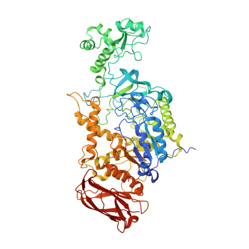
GtfC-type 4,6-α-glucanotransferase (α-GT) enzymes from Glycoside Hydrolase Family 70 (GH70) are of interest for the modification of starch into low-glycemic index food ingredients. Compared to the related GH70 GtfB-type α-GTs, found exclusively in lactic acid bacteria (LAB), GtfCs occur in non-LAB, share low sequence identity, lack circular permutation of the catalytic domain, and feature a single-segment auxiliary domain IV and auxiliary C-terminal domains. Despite these differences, the first crystal structure of a GtfC, GbGtfC-ΔC from Geobacillus 12AMOR1, and the first one representing a non-permuted GH70 enzyme, reveals high structural similarity in the core domains with most GtfBs, featuring a similar tunneled active site. We propose that GtfC (and related GtfD) enzymes evolved from starch-degrading α-amylases from GH13 by acquiring α-1,6 transglycosylation capabilities, before the events that resulted in circular permutation of the catalytic domain observed in other GH70 enzymes (glucansucrases, GtfB-type α-GTs). AlphaFold modeling and sequence alignments suggest that the GbGtfC structure represents the GtfC subfamily, although it has a so far unique alternating α-1,4/α-1,6 product specificity, likely determined by residues near acceptor binding subsites +1/+2.
- Biomolecular X-ray Crystallography, Groningen Biomolecular Sciences and Biotechnology Institute (GBB), University of Groningen, Nijenborgh 7, 9747 AG Groningen, The Netherlands.
Organizational Affiliation: