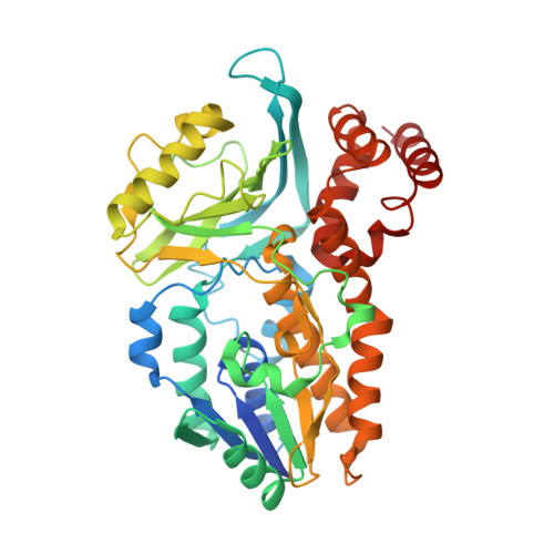
7Z4X

Crystal structure of Variovorax paradoxus indole monooxygenase (VpIndA1) in complex with FAD


Experimental Data Snapshot

  • Method: X-RAY DIFFRACTION
  • Resolution: 2.05 Å
  • R-Value Free: 0.213 
  • R-Value Work: 0.174 
  • R-Value Observed: 0.176 

Starting Model: in silico
View more details

wwPDB Validation   3D Report Full Report


Ligand Structure Quality Assessment 


This is version 1.2 of the entry. See complete history


Literature

Structural and Mechanistic Studies on Substrate and Stereoselectivity of the Indole Monooxygenase VpIndA1: New Avenues for Biocatalytic Epoxidations and Sulfoxidations.

Kratky, J.Eggerichs, D.Heine, T.Hofmann, S.Sowa, P.Weisse, R.H.Tischler, D.Strater, N.

(2023) Angew Chem Int Ed Engl 62: e202300657-e202300657

  • DOI: https://doi.org/10.1002/anie.202300657
  • Primary Citation of Related Structures:  
    7Z4X, 7Z94, 7Z97, 7Z98, 7Z99, 7ZCA

  • PubMed Abstract: 

    Flavoprotein monooxygenases are a versatile group of enzymes for biocatalytic transformations. Among these, group E monooxygenases (GEMs) catalyze enantioselective epoxidation and sulfoxidation reactions. Here, we describe the crystal structure of an indole monooxygenase from the bacterium Variovorax paradoxus EPS, a GEM designated as VpIndA1. Complex structures with substrates reveal productive binding modes that, in conjunction with force-field calculations and rapid mixing kinetics, reveal the structural basis of substrate and stereoselectivity. Structure-based redesign of the substrate cavity yielded variants with new substrate selectivity (for sulfoxidation of benzyl phenyl sulfide) or with greatly enhanced stereoselectivity (from 35.1 % to 99.8 % ee for production of (1S,2R)-indene oxide). This first determination of the substrate binding mode of GEMs combined with structure-function relationships opens the door for structure-based design of these powerful biocatalysts.


  • Organizational Affiliation

    Institute of Bioanalytical Chemistry, Leipzig University, Deutscher Platz 5, 04103, Leipzig, Germany.


Macromolecules
Find similar proteins by:  (by identity cutoff)  |  3D Structure
Entity ID: 1
MoleculeChains Sequence LengthOrganismDetailsImage
Putative dehydrogenase/oxygenase subunit (Flavoprotein)433Variovorax paradoxus EPSMutation(s): 0 
Gene Names: Varpa_4903
UniProt
Find proteins for E6V140 (Variovorax paradoxus (strain EPS))
Explore E6V140 
Go to UniProtKB:  E6V140
Entity Groups  
Sequence Clusters30% Identity50% Identity70% Identity90% Identity95% Identity100% Identity
UniProt GroupE6V140
Sequence Annotations
Expand
  • Reference Sequence
Small Molecules
Ligands 1 Unique
IDChains Name / Formula / InChI Key2D Diagram3D Interactions
FAD (Subject of Investigation/LOI)
Query on FAD

Download Ideal Coordinates CCD File 
B [auth A]FLAVIN-ADENINE DINUCLEOTIDE
C27 H33 N9 O15 P2
VWWQXMAJTJZDQX-UYBVJOGSSA-N
Experimental Data & Validation

Experimental Data

  • Method: X-RAY DIFFRACTION
  • Resolution: 2.05 Å
  • R-Value Free: 0.213 
  • R-Value Work: 0.174 
  • R-Value Observed: 0.176 
  • Space Group: P 32 2 1
Unit Cell:
Length ( Å )Angle ( ˚ )
a = 94.512α = 90
b = 94.512β = 90
c = 111.253γ = 120
Software Package:
Software NamePurpose
Aimlessdata scaling
PHENIXrefinement
PDB_EXTRACTdata extraction
XDSdata reduction
PHASERphasing

Structure Validation

View Full Validation Report


Ligand Structure Quality Assessment 


Entry History & Funding Information

Deposition Data


Funding OrganizationLocationGrant Number
Not funded--

Revision History  (Full details and data files)

  • Version 1.0: 2023-02-22
    Type: Initial release
  • Version 1.1: 2023-04-19
    Changes: Database references
  • Version 1.2: 2024-05-01
    Changes: Data collection, Refinement description