7Z4S | pdb_00007z4s

Crystal structure of SARS-CoV-2 Mpro in complex with cyclic peptide GM4 including unnatural amino acids.


Experimental Data Snapshot

  • Method: X-RAY DIFFRACTION
  • Resolution: 1.70 Å
  • R-Value Free: 
    0.234 (Depositor), 0.232 (DCC) 
  • R-Value Work: 
    0.198 (Depositor) 
  • R-Value Observed: 
    0.200 (Depositor) 

Starting Model: experimental
View more details

wwPDB Validation 3D Report Full Report

Validation slider image for 7Z4S

This is version 4.2 of the entry. See complete history

Literature

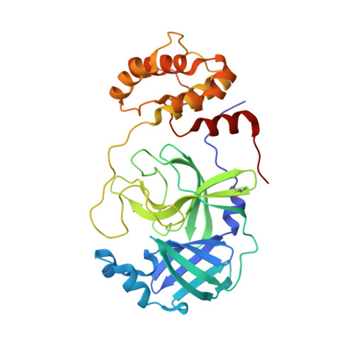
In vitro selection of macrocyclic peptide inhibitors containing cyclic gamma 2,4 -amino acids targeting the SARS-CoV-2 main protease.

Miura, T.Malla, T.R.Owen, C.D.Tumber, A.Brewitz, L.McDonough, M.A.Salah, E.Terasaka, N.Katoh, T.Lukacik, P.Strain-Damerell, C.Mikolajek, H.Walsh, M.A.Kawamura, A.Schofield, C.J.Suga, H.

(2023) Nat Chem 15: 998-1005

  • DOI: https://doi.org/10.1038/s41557-023-01205-1
  • Primary Citation Related Structures: 
    7Z4S

  • PubMed Abstract: 

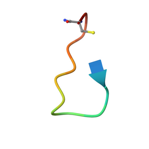
    γ-Amino acids can play important roles in the biological activities of natural products; however, the ribosomal incorporation of γ-amino acids into peptides is challenging. Here we report how a selection campaign employing a non-canonical peptide library containing cyclic γ 2,4 -amino acids resulted in the discovery of very potent inhibitors of the SARS-CoV-2 main protease (M pro ). Two kinds of cyclic γ 2,4 -amino acids, cis-3-aminocyclobutane carboxylic acid (γ 1 ) and (1R,3S)-3-aminocyclopentane carboxylic acid (γ 2 ), were ribosomally introduced into a library of thioether-macrocyclic peptides. One resultant potent M pro inhibitor (half-maximal inhibitory concentration = 50 nM), GM4, comprising 13 residues with γ 1 at the fourth position, manifests a 5.2 nM dissociation constant. An M pro :GM4 complex crystal structure reveals the intact inhibitor spans the substrate binding cleft. The γ 1 interacts with the S1' catalytic subsite and contributes to a 12-fold increase in proteolytic stability compared to its alanine-substituted variant. Knowledge of interactions between GM4 and M pro enabled production of a variant with a 5-fold increase in potency.


  • Organizational Affiliation
    • Department of Chemistry, Graduate School of Science, The University of Tokyo, Tokyo, Japan.

Macromolecule Content 

  • Total Structure Weight: 71.37 kDa 
  • Atom Count: 5,298 
  • Modeled Residue Count: 638 
  • Deposited Residue Count: 642 
  • Unique protein chains: 2

Macromolecules

Find similar proteins by:|  3D Structure
Entity ID: 1
MoleculeChains  Sequence LengthOrganismDetailsImage
3C-like proteinase nsp5
A, B
306Severe acute respiratory syndrome coronavirus 2Mutation(s): 0 
EC: 3.4.22.69
UniProt
Find proteins for P0DTC1 (Severe acute respiratory syndrome coronavirus 2)
Explore P0DTC1 
Go to UniProtKB:  P0DTC1
Entity Groups
Sequence Clusters30% Identity50% Identity70% Identity90% Identity95% Identity100% Identity
UniProt GroupP0DTC1
Sequence Annotations
Expand
Reference Sequence
Find similar proteins by:  Sequence   |   3D Structure  
Entity ID: 2
MoleculeChains  Sequence LengthOrganismDetailsImage
Macrocyclic peptide inhibitor
C, D
15synthetic constructMutation(s): 0 

Small Molecules

Ligands 4 Unique
IDChains Name / Formula / InChI Key2D Diagram3D Interactions
PGE

Query on PGE


Download:Ideal Coordinates CCD File
I [auth B]TRIETHYLENE GLYCOL
C6 H14 O4
ZIBGPFATKBEMQZ-UHFFFAOYSA-N
PEG

Query on PEG


Download:Ideal Coordinates CCD File
E [auth A],
G [auth B]
DI(HYDROXYETHYL)ETHER
C4 H10 O3
MTHSVFCYNBDYFN-UHFFFAOYSA-N
DMS

Query on DMS


Download:Ideal Coordinates CCD File
F [auth A]DIMETHYL SULFOXIDE
C2 H6 O S
IAZDPXIOMUYVGZ-UHFFFAOYSA-N
EDO

Query on EDO


Download:Ideal Coordinates CCD File
H [auth B],
J [auth B]
1,2-ETHANEDIOL
C2 H6 O2
LYCAIKOWRPUZTN-UHFFFAOYSA-N
Modified Residues  1 Unique
IDChains TypeFormula2D DiagramParent
CSO
Query on CSO
A, B
L-PEPTIDE LINKINGC3 H7 N O3 SCYS
DTY
Query on DTY
C, D
D-PEPTIDE LINKINGC9 H11 N O3

--

II7
Query on II7
C, D
L-PEPTIDE LINKINGC5 H9 N O2

--

Experimental Data & Validation

Experimental Data

  • Method: X-RAY DIFFRACTION
  • Resolution: 1.70 Å
  • R-Value Free:  0.234 (Depositor), 0.232 (DCC) 
  • R-Value Work:  0.198 (Depositor) 
  • R-Value Observed: 0.200 (Depositor) 
Space Group: P 1
Unit Cell:
Length ( Å )Angle ( ˚ )
a = 54.515α = 113.87
b = 56.112β = 110.53
c = 63.238γ = 90.92
Software Package:
Software NamePurpose
REFMACrefinement
xia2data reduction
Aimlessdata scaling
MOLREPphasing

Structure Validation

View Full Validation Report


Entry History 

& Funding Information

Deposition Data


Funding OrganizationLocationGrant Number
Not funded--

Revision History  (Full details and data files)

  • Version 1.0: 2023-03-22
    Type: Initial release
  • Version 2.0: 2023-05-24
    Changes: Atomic model, Derived calculations, Non-polymer description, Structure summary
  • Version 3.0: 2023-06-14
    Changes: Advisory, Atomic model, Author supporting evidence, Data collection, Database references, Derived calculations, Non-polymer description, Polymer sequence, Refinement description, Source and taxonomy, Structure summary
  • Version 3.1: 2023-07-19
    Changes: Data collection, Database references
  • Version 4.0: 2023-11-15
    Changes: Atomic model, Data collection
  • Version 4.1: 2024-02-07
    Changes: Refinement description
  • Version 4.2: 2024-10-16
    Changes: Structure summary