7Z2N | pdb_00007z2n

Tubulin-18-complex


Experimental Data Snapshot

  • Method: X-RAY DIFFRACTION
  • Resolution: 2.17 Å
  • R-Value Free: 
    0.224 (Depositor), 0.209 (DCC) 
  • R-Value Work: 
    0.189 (Depositor) 
  • R-Value Observed: 
    0.191 (Depositor) 

Starting Model: experimental
View more details

wwPDB Validation 3D Report Full Report

Validation slider image for 7Z2N

Ligand Structure Quality Assessment 


This is version 1.1 of the entry. See complete history

Literature

Novel fragment-derived colchicine-site binders as microtubule-destabilizing agents.

de la Roche, N.M.Muhlethaler, T.Di Martino, R.M.C.Ortega, J.A.Gioia, D.Roy, B.Prota, A.E.Steinmetz, M.O.Cavalli, A.

(2022) Eur J Med Chem 241: 114614-114614

  • DOI: https://doi.org/10.1016/j.ejmech.2022.114614
  • Primary Citation Related Structures: 
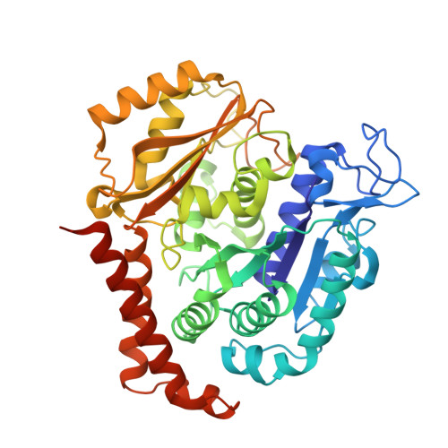
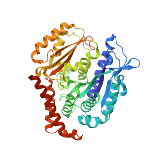
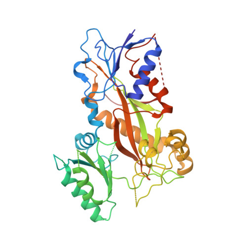
    7Z2N, 7Z2P

  • PubMed Abstract: 

    Microtubules (MTs) are dynamic filaments of the cytoskeleton, which are formed by the polymerization of their building block tubulin. Perturbation of MT dynamics by MT-targeting agents (MTAs) leads to cell cycle arrest or cell death, a strategy that is pursued in chemotherapy. We recently performed a combined computational and crystallographic fragment screening approach and identified several tubulin-binding fragments. Here, we sought to capitalize on this study with the aim to demonstrate that low affinity tubulin-binding fragments can indeed be used as valuable starting points for the development of active, lead-like antitubulin small molecules. To this end, we report on a new, rationally designed series of 2-aminobenzimidazole derivatives that destabilize MTs by binding tubulin at the colchicine-binding site (CBS). We applied a fragment growing strategy by combining X-ray crystallography and computer-aided drug design. Preliminary structure-activity-relationship studies afforded compound 18 that inhibits HeLa cell viability with a submicromolar activity (IC 50 of 0.9 μM). X-ray crystallography confirmed the compound pose in the CBS, while immunostaining experiments suggested a molecular mechanism of action alike classical CBS ligands with antimitotic and antitumor activity associated with MTs destabilization. This promising outcome underpins that our previously performed combined computational and crystallographic fragment screening approach provides promising starting points for developing new MTAs binding to the CBS of tubulin and, eventually, to further tubulin pockets.


  • Organizational Affiliation
    • Computational & Chemical Biology, Istituto Italiano di Tecnologia, Via Morego, 30, 16163, Genova, Italy; Department of Pharmacy and Biotechnology, Alma Mater Studiorum, University of Bologna, Via Belmeloro 6, 40126, Bologna, Italy.

Macromolecule Content 

  • Total Structure Weight: 264.85 kDa 
  • Atom Count: 18,229 
  • Modeled Residue Count: 2,193 
  • Deposited Residue Count: 2,319 
  • Unique protein chains: 4

Macromolecules

Find similar proteins by:|  3D Structure
Entity ID: 1
MoleculeChains  Sequence LengthOrganismDetailsImage
Tubulin alpha-1B chain
A, C
451Bos taurusMutation(s): 0 
EC: 3.6.5
UniProt
Find proteins for P81947 (Bos taurus)
Explore P81947 
Go to UniProtKB:  P81947
Entity Groups
Sequence Clusters30% Identity50% Identity70% Identity90% Identity95% Identity100% Identity
UniProt GroupP81947
Sequence Annotations
Expand
Reference Sequence
Find similar proteins by:|  3D Structure
Entity ID: 2
MoleculeChains  Sequence LengthOrganismDetailsImage
Tubulin beta-2B chain
B, D
445Bos taurusMutation(s): 0 
UniProt
Find proteins for Q6B856 (Bos taurus)
Explore Q6B856 
Go to UniProtKB:  Q6B856
Entity Groups
Sequence Clusters30% Identity50% Identity70% Identity90% Identity95% Identity100% Identity
UniProt GroupQ6B856
Sequence Annotations
Expand
Reference Sequence
Find similar proteins by:|  3D Structure
Entity ID: 3
MoleculeChains  Sequence LengthOrganismDetailsImage
Stathmin-4143Rattus norvegicusMutation(s): 0 
Gene Names: Stmn4
UniProt
Find proteins for P63043 (Rattus norvegicus)
Explore P63043 
Go to UniProtKB:  P63043
Entity Groups
Sequence Clusters30% Identity50% Identity70% Identity90% Identity95% Identity100% Identity
UniProt GroupP63043
Sequence Annotations
Expand
Reference Sequence
Find similar proteins by:|  3D Structure
Entity ID: 4
MoleculeChains  Sequence LengthOrganismDetailsImage
Tubulin beta-2B chain384Gallus gallusMutation(s): 0 
Gene Names: TTL

Small Molecules

Ligands 7 Unique
IDChains Name / Formula / InChI Key2D Diagram3D Interactions
GTP

Query on GTP


Download:Ideal Coordinates CCD File
G [auth A],
O [auth C]
GUANOSINE-5'-TRIPHOSPHATE
C10 H16 N5 O14 P3
XKMLYUALXHKNFT-UUOKFMHZSA-N
ACP

Query on ACP


Download:Ideal Coordinates CCD File
U [auth F]PHOSPHOMETHYLPHOSPHONIC ACID ADENYLATE ESTER
C11 H18 N5 O12 P3
UFZTZBNSLXELAL-IOSLPCCCSA-N
GDP

Query on GDP


Download:Ideal Coordinates CCD File
J [auth B],
R [auth D]
GUANOSINE-5'-DIPHOSPHATE
C10 H15 N5 O11 P2
QGWNDRXFNXRZMB-UUOKFMHZSA-N
IAZ
(Subject of Investigation/LOI)

Query on IAZ


Download:Ideal Coordinates CCD File
N [auth B]~{N}-(furan-2-ylmethyl)-6-phenoxy-1~{H}-benzimidazol-2-amine
C18 H15 N3 O2
KHBMCLOQYWKNDZ-UHFFFAOYSA-N
MES

Query on MES


Download:Ideal Coordinates CCD File
M [auth B]2-(N-MORPHOLINO)-ETHANESULFONIC ACID
C6 H13 N O4 S
SXGZJKUKBWWHRA-UHFFFAOYSA-N
CA

Query on CA


Download:Ideal Coordinates CCD File
I [auth A],
L [auth B],
Q [auth C],
T [auth E]
CALCIUM ION
Ca
BHPQYMZQTOCNFJ-UHFFFAOYSA-N
MG

Query on MG


Download:Ideal Coordinates CCD File
H [auth A],
K [auth B],
P [auth C],
S [auth D],
V [auth F]
MAGNESIUM ION
Mg
JLVVSXFLKOJNIY-UHFFFAOYSA-N

Experimental Data & Validation

Experimental Data

  • Method: X-RAY DIFFRACTION
  • Resolution: 2.17 Å
  • R-Value Free:  0.224 (Depositor), 0.209 (DCC) 
  • R-Value Work:  0.189 (Depositor) 
  • R-Value Observed: 0.191 (Depositor) 
Space Group: P 21 21 21
Unit Cell:
Length ( Å )Angle ( ˚ )
a = 105.016α = 90
b = 157.703β = 90
c = 180.154γ = 90
Software Package:
Software NamePurpose
PHENIXrefinement
PDB_EXTRACTdata extraction
XDSdata reduction
XSCALEdata scaling
PHENIXphasing

Structure Validation

View Full Validation Report


Ligand Structure Quality Assessment 


Entry History 

& Funding Information

Deposition Data


Funding OrganizationLocationGrant Number
Swiss National Science FoundationSwitzerland31003A_166608
European CommissionEuropean UnionID239047 NEON

Revision History  (Full details and data files)

  • Version 1.0: 2022-08-31
    Type: Initial release
  • Version 1.1: 2024-01-31
    Changes: Data collection, Refinement description