7Z15 | pdb_00007z15

E. coli C-P lyase bound to a PhnK/PhnL dual ABC dimer and ADP + Pi


Experimental Data Snapshot

  • Method: ELECTRON MICROSCOPY
  • Resolution: 1.93 Å
  • Aggregation State: PARTICLE 
  • Reconstruction Method: SINGLE PARTICLE 

Starting Model: experimental
View more details

wwPDB Validation 3D Report Full Report

Validation slider image for 7Z15

This is version 1.2 of the entry. See complete history

Literature

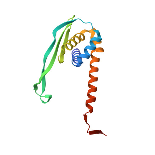
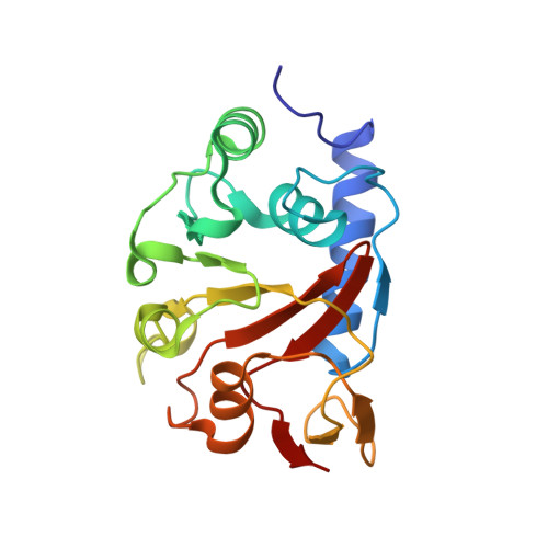
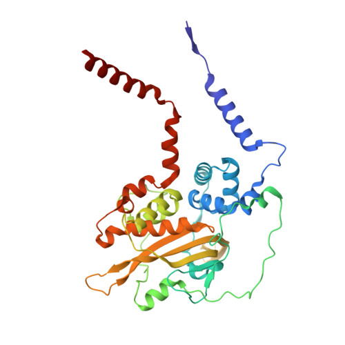
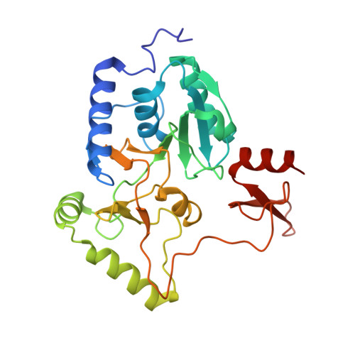
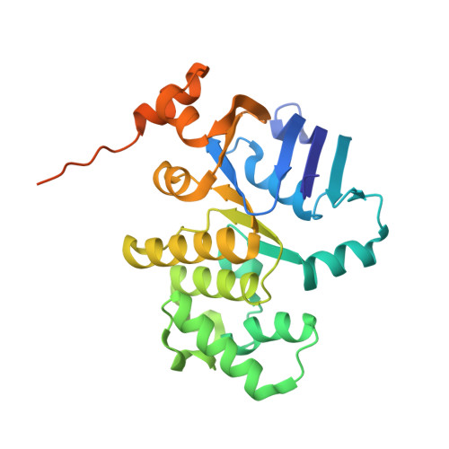
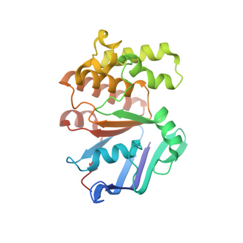
Structural remodelling of the carbon-phosphorus lyase machinery by a dual ABC ATPase.

Amstrup, S.K.Ong, S.C.Sofos, N.Karlsen, J.L.Skjerning, R.B.Boesen, T.Enghild, J.J.Hove-Jensen, B.Brodersen, D.E.

(2023) Nat Commun 14: 1001-1001

  • DOI: https://doi.org/10.1038/s41467-023-36604-y
  • Primary Citation Related Structures: 
    7Z15, 7Z16, 7Z17, 7Z18, 7Z19

  • PubMed Abstract: 

    In Escherichia coli, the 14-cistron phn operon encoding carbon-phosphorus lyase allows for utilisation of phosphorus from a wide range of stable phosphonate compounds containing a C-P bond. As part of a complex, multi-step pathway, the PhnJ subunit was shown to cleave the C-P bond via a radical mechanism, however, the details of the reaction could not immediately be reconciled with the crystal structure of a 220 kDa PhnGHIJ C-P lyase core complex, leaving a significant gap in our understanding of phosphonate breakdown in bacteria. Here, we show using single-particle cryogenic electron microscopy that PhnJ mediates binding of a double dimer of the ATP-binding cassette proteins, PhnK and PhnL, to the core complex. ATP hydrolysis induces drastic structural remodelling leading to opening of the core complex and reconfiguration of a metal-binding and putative active site located at the interface between the PhnI and PhnJ subunits.


  • Organizational Affiliation
    • Department of Molecular Biology and Genetics, Aarhus University, Universitetsbyen 81, DK-8000, Aarhus C, Denmark.

Macromolecule Content 

  • Total Structure Weight: 333.34 kDa 
  • Atom Count: 23,755 
  • Modeled Residue Count: 2,888 
  • Deposited Residue Count: 2,992 
  • Unique protein chains: 6

Macromolecules

Find similar proteins by:|  3D Structure
Entity ID: 1
MoleculeChains  Sequence LengthOrganismDetailsImage
Alpha-D-ribose 1-methylphosphonate 5-triphosphate synthase subunit PhnG
A, E
150Escherichia coliMutation(s): 0 
Gene Names: phnGb4101JW4062
EC: 2.7.8.37
UniProt
Find proteins for P16685 (Escherichia coli (strain K12))
Explore P16685 
Go to UniProtKB:  P16685
Entity Groups
Sequence Clusters30% Identity50% Identity70% Identity90% Identity95% Identity100% Identity
UniProt GroupP16685
Sequence Annotations
Expand
Reference Sequence
Find similar proteins by:|  3D Structure
Entity ID: 2
MoleculeChains  Sequence LengthOrganismDetailsImage
Alpha-D-ribose 1-methylphosphonate 5-triphosphate synthase subunit PhnH
B, F
194Escherichia coliMutation(s): 0 
Gene Names: phnHb4100JW4061
EC: 2.7.8.37
UniProt
Find proteins for P16686 (Escherichia coli (strain K12))
Explore P16686 
Go to UniProtKB:  P16686
Entity Groups
Sequence Clusters30% Identity50% Identity70% Identity90% Identity95% Identity100% Identity
UniProt GroupP16686
Sequence Annotations
Expand
Reference Sequence
Find similar proteins by:|  3D Structure
Entity ID: 3
MoleculeChains  Sequence LengthOrganismDetailsImage
Alpha-D-ribose 1-methylphosphonate 5-triphosphate synthase subunit PhnI
C, G
354Escherichia coliMutation(s): 0 
Gene Names: phnIb4099JW4060
EC: 2.7.8.37
UniProt
Find proteins for P16687 (Escherichia coli (strain K12))
Explore P16687 
Go to UniProtKB:  P16687
Entity Groups
Sequence Clusters30% Identity50% Identity70% Identity90% Identity95% Identity100% Identity
UniProt GroupP16687
Sequence Annotations
Expand
Reference Sequence
Find similar proteins by:|  3D Structure
Entity ID: 4
MoleculeChains  Sequence LengthOrganismDetailsImage
Alpha-D-ribose 1-methylphosphonate 5-phosphate C-P lyase
D, H
281Escherichia coliMutation(s): 0 
Gene Names: phnJb4098JW4059
EC: 4.7.1.1
UniProt
Find proteins for P16688 (Escherichia coli (strain K12))
Explore P16688 
Go to UniProtKB:  P16688
Entity Groups
Sequence Clusters30% Identity50% Identity70% Identity90% Identity95% Identity100% Identity
UniProt GroupP16688
Sequence Annotations
Expand
Reference Sequence
Find similar proteins by:|  3D Structure
Entity ID: 5
MoleculeChains  Sequence LengthOrganismDetailsImage
Putative phosphonates utilization ATP-binding protein PhnK
I, J
291Escherichia coliMutation(s): 0 
Gene Names: phnKb4097JW5727
UniProt
Find proteins for P16678 (Escherichia coli (strain K12))
Explore P16678 
Go to UniProtKB:  P16678
Entity Groups
Sequence Clusters30% Identity50% Identity70% Identity90% Identity95% Identity100% Identity
UniProt GroupP16678
Sequence Annotations
Expand
Reference Sequence
Find similar proteins by:|  3D Structure
Entity ID: 6
MoleculeChains  Sequence LengthOrganismDetailsImage
Alpha-D-ribose 1-methylphosphonate 5-triphosphate synthase subunit PhnL
K, L
226Escherichia coliMutation(s): 0 
Gene Names: phnLb4096JW4057
EC: 2.7.8.37
UniProt
Find proteins for P16679 (Escherichia coli (strain K12))
Explore P16679 
Go to UniProtKB:  P16679
Entity Groups
Sequence Clusters30% Identity50% Identity70% Identity90% Identity95% Identity100% Identity
UniProt GroupP16679
Sequence Annotations
Expand
Reference Sequence

Small Molecules

Ligands 6 Unique
IDChains Name / Formula / InChI Key2D Diagram3D Interactions
ATP

Query on ATP


Download:Ideal Coordinates CCD File
AA [auth L],
Y [auth K]
ADENOSINE-5'-TRIPHOSPHATE
C10 H16 N5 O13 P3
ZKHQWZAMYRWXGA-KQYNXXCUSA-N
ADP
(Subject of Investigation/LOI)

Query on ADP


Download:Ideal Coordinates CCD File
S [auth I],
V [auth J]
ADENOSINE-5'-DIPHOSPHATE
C10 H15 N5 O10 P2
XTWYTFMLZFPYCI-KQYNXXCUSA-N
I9X
(Subject of Investigation/LOI)

Query on I9X


Download:Ideal Coordinates CCD File
O [auth D],
R [auth H]
alpha-D-ribose-1,2-cyclic-phosphate-5-phosphate
C5 H10 O10 P2
OXGUIUWFXGIWNM-TXICZTDVSA-N
PO4
(Subject of Investigation/LOI)

Query on PO4


Download:Ideal Coordinates CCD File
T [auth I],
W [auth J]
PHOSPHATE ION
O4 P
NBIIXXVUZAFLBC-UHFFFAOYSA-K
ZN
(Subject of Investigation/LOI)

Query on ZN


Download:Ideal Coordinates CCD File
M [auth C],
N [auth D],
P [auth G],
Q [auth H]
ZINC ION
Zn
PTFCDOFLOPIGGS-UHFFFAOYSA-N
MG

Query on MG


Download:Ideal Coordinates CCD File
BA [auth L],
U [auth I],
X [auth J],
Z [auth K]
MAGNESIUM ION
Mg
JLVVSXFLKOJNIY-UHFFFAOYSA-N

Experimental Data & Validation

Experimental Data

  • Method: ELECTRON MICROSCOPY
  • Resolution: 1.93 Å
  • Aggregation State: PARTICLE 
  • Reconstruction Method: SINGLE PARTICLE 
EM Software:
TaskSoftware PackageVersion
RECONSTRUCTIONcryoSPARC3.2
MODEL REFINEMENTPHENIX

Structure Validation

View Full Validation Report


Entry History 

& Funding Information

Deposition Data


Funding OrganizationLocationGrant Number
Novo Nordisk FoundationDenmarkNNF18OC0030646

Revision History  (Full details and data files)

  • Version 1.0: 2022-06-22
    Type: Initial release
  • Version 1.1: 2023-03-08
    Changes: Database references, Refinement description
  • Version 1.2: 2024-07-17
    Changes: Data collection, Refinement description