7YVB

Aplysia californica FaNaC in ligand bound state


Experimental Data Snapshot

  • Method: ELECTRON MICROSCOPY
  • Resolution: 2.90 Å
  • Resolution: 2.90 Å
  • Resolution: 2.90 Å
  • Resolution: 2.90 Å
  • Resolution: 2.90 Å
  • Resolution: 2.90 Å
  • Resolution: 2.90 Å
  • Resolution: 2.90 Å
  • Resolution: 2.90 Å
  • Resolution: 2.90 Å
  • Resolution: 2.90 Å
  • Resolution: 2.90 Å
  • Resolution: 3.00 Å
  • Resolution: 2.90 Å
  • Resolution: 2.90 Å
  • Resolution: 2.90 Å
  • Aggregation State: PARTICLE 
  • Reconstruction Method: SINGLE PARTICLE 

wwPDB Validation   3D Report Full Report


This is version 1.2 of the entry. See complete history


Literature

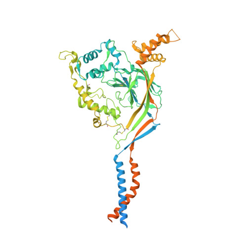
Structure and mechanism of a neuropeptide-activated channel in the ENaC/DEG superfamily.

Liu, F.Dang, Y.Li, L.Feng, H.Li, J.Wang, H.Zhang, X.Zhang, Z.Ye, S.Tian, Y.Chen, Q.

(2023) Nat Chem Biol 19: 1276-1285

  • DOI: https://doi.org/10.1038/s41589-023-01401-7
  • Primary Citation of Related Structures:  
    7YVB, 7YVC

  • PubMed Abstract: 

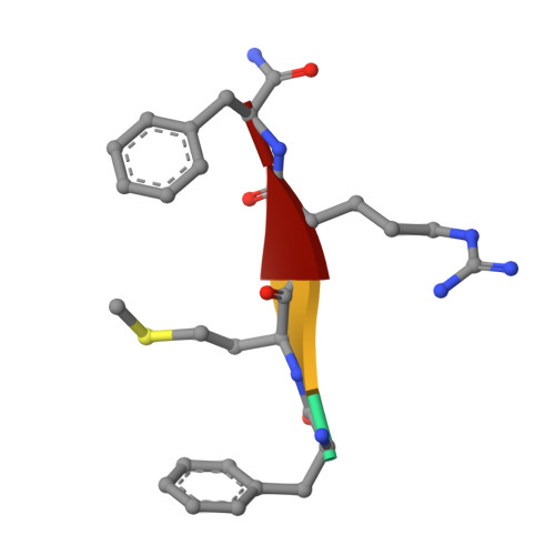
    Phe-Met-Arg-Phe-amide (FMRFamide)-activated sodium channels (FaNaCs) are a family of channels activated by the neuropeptide FMRFamide, and, to date, the underlying ligand gating mechanism remains unknown. Here we present the high-resolution cryo-electron microscopy structures of Aplysia californica FaNaC in both apo and FMRFamide-bound states. AcFaNaC forms a chalice-shaped trimer and possesses several notable features, including two FaNaC-specific insertion regions, a distinct finger domain and non-domain-swapped transmembrane helix 2 in the transmembrane domain (TMD). One FMRFamide binds to each subunit in a cleft located in the top-most region of the extracellular domain, with participation of residues from the neighboring subunit. Bound FMRFamide adopts an extended conformation. FMRFamide binds tightly to A. californica FaNaC in an N terminus-in manner, which causes collapse of the binding cleft and induces large local conformational rearrangements. Such conformational changes are propagated downward toward the TMD via the palm domain, possibly resulting in outward movement of the TMD and dilation of the ion conduction pore.


  • Organizational Affiliation

    State Key Laboratory of Conservation and Utilization of Bio-Resources in Yunnan and Center for Life Sciences, School of Life Sciences, Yunnan University, Kunming, China.


Macromolecules
Find similar proteins by:  (by identity cutoff)  |  3D Structure
Entity ID: 1
MoleculeChains Sequence LengthOrganismDetailsImage
FMRFamide-gated Na+ channel
A, B, C
679Aplysia californicaMutation(s): 0 
Membrane Entity: Yes 
UniProt
Find proteins for Q4TZI8 (Aplysia californica)
Explore Q4TZI8 
Go to UniProtKB:  Q4TZI8
Entity Groups  
Sequence Clusters30% Identity50% Identity70% Identity90% Identity95% Identity100% Identity
UniProt GroupQ4TZI8
Sequence Annotations
Expand
  • Reference Sequence

Find similar proteins by:  Sequence   |   3D Structure  

Entity ID: 2
MoleculeChains Sequence LengthOrganismDetailsImage
Phe-Met-Arg-Phe-amideD [auth P],
E [auth Q],
F [auth R]
4synthetic constructMutation(s): 0 
Sequence Annotations
Expand
  • Reference Sequence
Oligosaccharides

Help

Entity ID: 3
MoleculeChains Length2D Diagram Glycosylation3D Interactions
2-acetamido-2-deoxy-beta-D-glucopyranose-(1-4)-2-acetamido-2-deoxy-beta-D-glucopyranose
G [auth D],
H [auth E],
I [auth F],
J [auth G],
K [auth H],
G [auth D],
H [auth E],
I [auth F],
J [auth G],
K [auth H],
L [auth I],
M [auth J],
N [auth K],
O [auth L],
P [auth M],
Q [auth N],
R [auth O]
2N-Glycosylation
Glycosylation Resources
GlyTouCan:  G42666HT
GlyCosmos:  G42666HT
GlyGen:  G42666HT
Experimental Data & Validation

Experimental Data

  • Method: ELECTRON MICROSCOPY
  • Resolution: 2.90 Å
  • Resolution: 2.90 Å
  • Resolution: 2.90 Å
  • Resolution: 2.90 Å
  • Resolution: 2.90 Å
  • Resolution: 2.90 Å
  • Resolution: 2.90 Å
  • Resolution: 2.90 Å
  • Resolution: 2.90 Å
  • Resolution: 2.90 Å
  • Resolution: 2.90 Å
  • Resolution: 2.90 Å
  • Resolution: 3.00 Å
  • Resolution: 2.90 Å
  • Resolution: 2.90 Å
  • Resolution: 2.90 Å
  • Aggregation State: PARTICLE 
  • Reconstruction Method: SINGLE PARTICLE 

Structure Validation

View Full Validation Report


Entry History & Funding Information

Deposition Data


Funding OrganizationLocationGrant Number
National Natural Science Foundation of China (NSFC)China32071202
National Natural Science Foundation of China (NSFC)China32171201

Revision History  (Full details and data files)

  • Version 1.0: 2023-08-09
    Type: Initial release
  • Version 1.1: 2023-08-30
    Changes: Data collection, Database references
  • Version 1.2: 2023-10-11
    Changes: Database references