


Human langerin carbohydrate recognition domain in complex with an alpha-mannoside ligand
Wangkanont, K.To be published.
Experimental Data Snapshot
Starting Model: experimental
View more details
Entity ID: 1 | |||||
|---|---|---|---|---|---|
| Molecule | Chains | Sequence Length | Organism | Details | Image |
| CD207 molecule | 156 | Homo sapiens | Mutation(s): 0 Gene Names: CD207 | 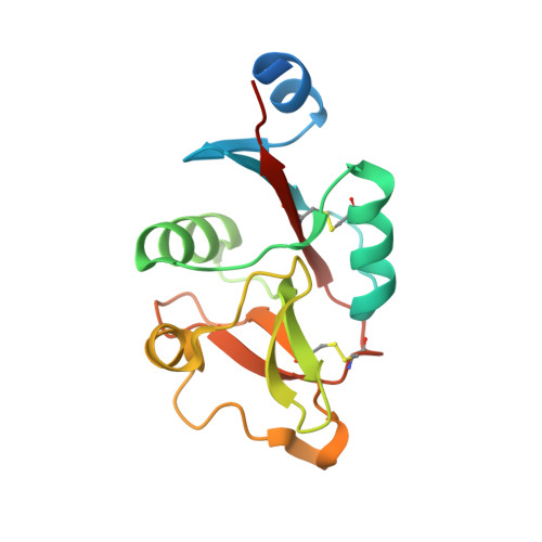 | |
UniProt & NIH Common Fund Data Resources | |||||
PHAROS: Q9UJ71 GTEx: ENSG00000116031 | |||||
Entity Groups | |||||
| Sequence Clusters | 30% Identity50% Identity70% Identity90% Identity95% Identity100% Identity | ||||
| UniProt Group | Q9UJ71 | ||||
Sequence AnnotationsExpand | |||||
Reference Sequence | |||||
Entity ID: 2 | |||||
|---|---|---|---|---|---|
| Molecule | Chains | Sequence Length | Organism | Details | Image |
| SER-TRP | 2 | unidentified | Mutation(s): 0 | 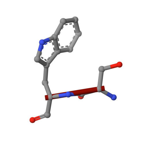 | |
| Ligands 4 Unique | |||||
|---|---|---|---|---|---|
| ID | Chains | Name / Formula / InChI Key | 2D Diagram | 3D Interactions | |
| JMI (Subject of Investigation/LOI) Download:Ideal Coordinates CCD File | G [auth A], J [auth B], N [auth C], R [auth D] | ~{N}-[2-[4-[(2~{R},3~{S},4~{S},5~{S},6~{R})-6-(hydroxymethyl)-3,4,5-tris(oxidanyl)oxan-2-yl]oxyphenyl]ethyl]ethanamide C16 H23 N O7 JVYCDFJBIOIPDL-OWYFMNJBSA-N |  | ||
| TRP Download:Ideal Coordinates CCD File | L [auth C], P [auth D] | TRYPTOPHAN C11 H12 N2 O2 QIVBCDIJIAJPQS-VIFPVBQESA-N |  | ||
| CA Download:Ideal Coordinates CCD File | F [auth A], I [auth B], M [auth C], Q [auth D] | CALCIUM ION Ca BHPQYMZQTOCNFJ-UHFFFAOYSA-N |  | ||
| CL Download:Ideal Coordinates CCD File | H [auth A], K [auth B], O [auth C], S [auth D] | CHLORIDE ION Cl VEXZGXHMUGYJMC-UHFFFAOYSA-M |  | ||
| Length ( Å ) | Angle ( ˚ ) |
|---|---|
| a = 80.03 | α = 90 |
| b = 80.03 | β = 90 |
| c = 90.07 | γ = 90 |
| Software Name | Purpose |
|---|---|
| PHENIX | refinement |
| Aimless | data scaling |
| MOSFLM | data reduction |
| PHASER | phasing |
| Funding Organization | Location | Grant Number |
|---|---|---|
| Chulalongkorn University | Thailand | -- |


