Structural insights into FSP1 catalysis and ferroptosis inhibition.
Lv, Y., Liang, C., Sun, Q., Zhu, J., Xu, H., Li, X., Li, Y.Y., Wang, Q., Yuan, H., Chu, B., Zhu, D.(2023) Nat Commun 14: 5933-5933
- PubMed: 37739943 Search on PubMedSearch on PubMed Central
- DOI: https://doi.org/10.1038/s41467-023-41626-7
- Primary Citation Related Structures:
7XPI, 7YTL - PubMed Abstract:
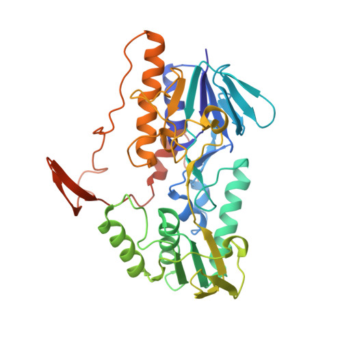
Ferroptosis suppressor protein 1 (FSP1, also known as AIMF2, AMID or PRG3) is a recently identified glutathione-independent ferroptosis suppressor 1-3 , but its underlying structural mechanism remains unknown. Here we report the crystal structures of Gallus gallus FSP1 in its substrate-free and ubiquinone-bound forms. The structures reveal a FAD-binding domain and a NAD(P)H-binding domain, both of which are shared with AIF and NADH oxidoreductases 4-9 , and a characteristic carboxy-terminal domain as well. We demonstrate that the carboxy-terminal domain is crucial for the catalytic activity and ferroptosis inhibition of FSP1 by mediating the functional dimerization of FSP1, and the formation of two active sites located on two sides of FAD, which are responsible for ubiquinone reduction and a unique FAD hydroxylation respectively. We also identify that FSP1 can catalyze the production of H 2 O 2 and the conversion of FAD to 6-hydroxy-FAD in the presence of oxygen and NAD(P)H in vitro, and 6-hydroxy-FAD directly inhibits ferroptosis in cells. Together, these findings further our understanding on the catalytic and ferroptosis suppression mechanisms of FSP1 and establish 6-hydroxy-FAD as an active cofactor in FSP1 and a potent radical-trapping antioxidant in ferroptosis inhibition.
- Department of Biochemistry and Molecular Biology, School of Basic Medical Sciences, Cheeloo College of Medicine, Shandong University, Jinan, 250012, China.
Organizational Affiliation: