7YRO

Crystal structure of mango fucosyltransferase 13


Experimental Data Snapshot

  • Method: X-RAY DIFFRACTION
  • Resolution: 2.42 Å
  • R-Value Free: 0.248 
  • R-Value Work: 0.210 
  • R-Value Observed: 0.211 

wwPDB Validation   3D Report Full Report


Ligand Structure Quality Assessment 


This is version 1.1 of the entry. See complete history


Literature

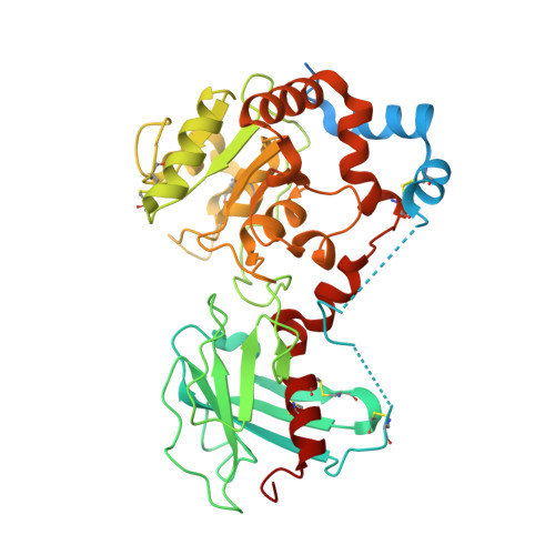
Crystal structure of mango alpha 1,3/ alpha 1,4-fucosyltransferase elucidates unique elements that regulate Lewis A-dominant oligosaccharide assembly.

Okada, T.Teramoto, T.Ihara, H.Ikeda, Y.Kakuta, Y.

(2024) Glycobiology 34

  • DOI: https://doi.org/10.1093/glycob/cwae015
  • Primary Citation of Related Structures:  
    7YRO

  • PubMed Abstract: 

    In various organisms, α1,3/α1,4-fucosyltransferases (CAZy GT10 family enzymes) mediate the assembly of type I (Galβ1,3GlcNAc) and/or type II (Galβ1,4GlcNAc)-based Lewis structures that are widely distributed in glycoconjugates. Unlike enzymes of other species, plant orthologues show little fucosyltransferase activity for type II-based glycans and predominantly catalyze the assembly of the Lewis A structure [Galβ1,3(Fucα1,4)GlcNAc] on the type I disaccharide unit of their substrates. However, the structural basis underlying this unique substrate selectivity remains elusive. In this study, we investigated the structure-function relationship of MiFUT13A, a mango α1,3/α1,4-fucosyltransferase. The prepared MiFUT13A displayed distinct α1,4-fucosyltransferase activity. Consistent with the enzymatic properties of this molecule, X-ray crystallography revealed that this enzyme has a typical GT-B fold-type structure containing a set of residues that are responsible for its SN2-like catalysis. Site-directed mutagenesis and molecular docking analyses proposed a rational binding mechanism for type I oligosaccharides. Within the catalytic cleft, the pocket surrounding Trp121 serves as a binding site, anchoring the non-reducing terminal β1,3-galactose that belongs to the type I disaccharide unit. Furthermore, Glu177 was postulated to function as a general base catalyst through its interaction with the 4-hydroxy group of the acceptor N-acetylglucosamine residue. Adjacent residues, specifically Thr120, Thr157 and Asp175 were speculated to assist in binding of the reducing terminal residues. Intriguingly, these structural elements were not fully conserved in mammalian orthologue which also shows predominant α1,4-fucosyltransferase activity. In conclusion, we have proposed that MiFUT13A generates the Lewis A structure on type I glycans through a distinct mechanism, divergent from that of mammalian enzymes.


  • Organizational Affiliation

    Division of Molecular Cell Biology, Department of Biomolecular Sciences, Faculty of Medicine, Saga University, 5-1-1 Nabeshima, Saga 849-8501, Japan.


Macromolecules
Find similar proteins by:  (by identity cutoff)  |  3D Structure
Entity ID: 1
MoleculeChains Sequence LengthOrganismDetailsImage
Fucosyltransferase
A, B, C, D, E
A, B, C, D, E, F, G, H
410Mangifera indicaMutation(s): 0 
Gene Names: FUT13
EC: 2.4.1
UniProt
Find proteins for A0A292GKJ7 (Mangifera indica)
Explore A0A292GKJ7 
Go to UniProtKB:  A0A292GKJ7
Entity Groups  
Sequence Clusters30% Identity50% Identity70% Identity90% Identity95% Identity100% Identity
UniProt GroupA0A292GKJ7
Sequence Annotations
Expand
  • Reference Sequence
Small Molecules
Ligands 5 Unique
IDChains Name / Formula / InChI Key2D Diagram3D Interactions
NAG (Subject of Investigation/LOI)
Query on NAG

Download Ideal Coordinates CCD File 
AA [auth D],
GA [auth E],
M [auth B],
PA [auth F],
R [auth C]
2-acetamido-2-deoxy-beta-D-glucopyranose
C8 H15 N O6
OVRNDRQMDRJTHS-FMDGEEDCSA-N
PPV (Subject of Investigation/LOI)
Query on PPV

Download Ideal Coordinates CCD File 
FA [auth E]
L [auth B]
LA [auth F]
Q [auth C]
RA [auth G]
FA [auth E],
L [auth B],
LA [auth F],
Q [auth C],
RA [auth G],
UA [auth H],
Z [auth D]
PYROPHOSPHATE
H4 O7 P2
XPPKVPWEQAFLFU-UHFFFAOYSA-N
PO4 (Subject of Investigation/LOI)
Query on PO4

Download Ideal Coordinates CCD File 
K [auth A]PHOSPHATE ION
O4 P
NBIIXXVUZAFLBC-UHFFFAOYSA-K
GOL (Subject of Investigation/LOI)
Query on GOL

Download Ideal Coordinates CCD File 
BA [auth D]
CA [auth D]
DA [auth D]
EA [auth D]
HA [auth E]
BA [auth D],
CA [auth D],
DA [auth D],
EA [auth D],
HA [auth E],
I [auth A],
JA [auth E],
KA [auth E],
MA [auth F],
N [auth B],
NA [auth F],
O [auth B],
OA [auth F],
S [auth C],
SA [auth G],
T [auth C],
U [auth C],
VA [auth H],
W [auth C],
X [auth C],
Y [auth C]
GLYCEROL
C3 H8 O3
PEDCQBHIVMGVHV-UHFFFAOYSA-N
ACY (Subject of Investigation/LOI)
Query on ACY

Download Ideal Coordinates CCD File 
IA [auth E]
J [auth A]
P [auth B]
QA [auth F]
TA [auth G]
IA [auth E],
J [auth A],
P [auth B],
QA [auth F],
TA [auth G],
V [auth C],
WA [auth H]
ACETIC ACID
C2 H4 O2
QTBSBXVTEAMEQO-UHFFFAOYSA-N
Experimental Data & Validation

Experimental Data

  • Method: X-RAY DIFFRACTION
  • Resolution: 2.42 Å
  • R-Value Free: 0.248 
  • R-Value Work: 0.210 
  • R-Value Observed: 0.211 
  • Space Group: P 21 21 21
Unit Cell:
Length ( Å )Angle ( ˚ )
a = 86.7α = 90
b = 163.61β = 90
c = 274.9γ = 90
Software Package:
Software NamePurpose
PHENIXrefinement
XDSdata reduction
XDSdata scaling
PHASERphasing

Structure Validation

View Full Validation Report


Ligand Structure Quality Assessment 


Entry History & Funding Information

Deposition Data


Funding OrganizationLocationGrant Number
Japan Society for the Promotion of Science (JSPS)Japan20K06019

Revision History  (Full details and data files)

  • Version 1.0: 2023-08-16
    Type: Initial release
  • Version 1.1: 2024-06-19
    Changes: Database references