The crystal structure of Vibrio cholerae (6-4) photolyase reveals interactions with cofactors and a DNA-binding region.
Cakilkaya, B., Kavakli, I.H., DeMirci, H.(2023) J Biological Chem 299: 102794-102794
- PubMed: 36528063 Search on PubMedSearch on PubMed Central
- DOI: https://doi.org/10.1016/j.jbc.2022.102794
- Primary Citation Related Structures:
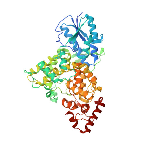
7YKN - PubMed Abstract:
Photolyases (PLs) reverse UV-induced DNA damage using blue light as an energy source. Of these PLs, (6-4) PLs repair (6-4)-lesioned photoproducts. We recently identified a gene from Vibrio cholerae (Vc) encoding a (6-4) PL, but structural characterization is needed to elucidate specific interactions with the chromophore cofactors. Here, we determined the crystal structure of Vc (6-4) PL at 2.5 Å resolution. Our high-resolution structure revealed that the two well-known cofactors, flavin adenine dinucleotide and the photoantenna 6,7-dimethyl 8-ribityl-lumazin (DMRL), stably interact with an α-helical and an α/β domain, respectively. Additionally, the structure has a third cofactor with distinct electron clouds corresponding to a [4Fe-4S] cluster. Moreover, we identified that Asp106 makes a hydrogen bond with water and DMRL, which indicates further stabilization of the photoantenna DMRL within Vc (6-4) PL. Further analysis of the Vc (6-4) PL structure revealed a possible region responsible for DNA binding. The region located between residues 478 to 484 may bind the lesioned DNA, with Arg483 potentially forming a salt bridge with DNA to stabilize further the interaction of Vc (6-4) PL with its substrate. Our comparative analysis revealed that the DNA lesion could not bind to the Vc (6-4) PL in a similar fashion to the Drosophila melanogaster (Dm, (6-4)) PL without a significant conformational change of the protein. The 23rd helix of the bacterial (6-4) PLs seems to have remarkable plasticity, and conformational changes facilitate DNA binding. In conclusion, our structure provides further insight into DNA repair by a (6-4) PL containing three cofactors.
- Department of Molecular Biology and Genetics, Koc University, Istanbul, Turkey.
Organizational Affiliation: