7YHJ | pdb_00007yhj

Effector binding domain of LysR-Type transcription factor LrhA from E. coli


Experimental Data Snapshot

  • Method: X-RAY DIFFRACTION
  • Resolution: 3.24 Å
  • R-Value Free: 
    0.338 (Depositor), 0.330 (DCC) 
  • R-Value Work: 
    0.321 (Depositor), 0.320 (DCC) 
  • R-Value Observed: 
    0.322 (Depositor) 

Starting Model: experimental
View more details

wwPDB Validation   3D Report Full Report


This is version 1.2 of the entry. See complete history


Literature

Oligomerization-Dependent Regulation of LrhA Controls Bacterial Flagellar Biosynthesis.

Niu, B.Kikkawa, M.Jiang, X.

(2026) J Mol Biology 438: 169682-169682

  • DOI: https://doi.org/10.1016/j.jmb.2026.169682
  • Primary Citation of Related Structures:  
    22XO, 7YHJ

  • PubMed Abstract: 

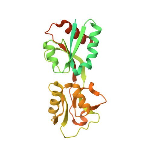
    LysR-type transcriptional regulators (LTTRs) are a diverse family of proteins that regulate various cellular processes, including motility in bacteria. In Escherichia coli, the LTTR LrhA represses flagellar biosynthesis by inhibiting the flhDC operon. However, the structural basis underlying this regulation has remained unclear. Here, we determined both a high-resolution crystal structure and a cryo-EM reconstruction of LrhA, revealing a predominant and stable tetrameric organization with pronounced structural variability in its effector-binding region. Structural and biochemical analyses demonstrate that mutations in these variable regions perturb the oligomeric equilibrium of LrhA, shifting the balance between tetrameric and dimeric species. This shift correlates with enhanced DNA binding affinity and stronger repression of the flhDC promoter. While ligand binding may similarly modulate LrhA activity, our data primarily support a model in which alterations in oligomeric state mediated by the variable regions regulate LrhA function. Together, these findings provide a structural framework for understanding how LrhA controls bacterial motility and offer broader insights into oligomerization-based regulation within the LTTR family.


  • Organizational Affiliation
    • Department of Cell Biology and Anatomy, Graduate School of Medicine, The University of Tokyo, Tokyo 113-0033, Japan.

Macromolecules
Find similar proteins by:  (by identity cutoff)  |  3D Structure
Entity ID: 1
MoleculeChains Sequence LengthOrganismDetailsImage
Probable HTH-type transcriptional regulator LrhA
A, B, C, D, E
A, B, C, D, E, F, G, H
319Escherichia coliMutation(s): 0 
Gene Names: lrhAgenRb2289JW2284
UniProt
Find proteins for P36771 (Escherichia coli (strain K12))
Explore P36771 
Go to UniProtKB:  P36771
Entity Groups  
Sequence Clusters30% Identity50% Identity70% Identity90% Identity95% Identity100% Identity
UniProt GroupP36771
Sequence Annotations
Expand
  • Reference Sequence
Experimental Data & Validation

Experimental Data

  • Method: X-RAY DIFFRACTION
  • Resolution: 3.24 Å
  • R-Value Free:  0.338 (Depositor), 0.330 (DCC) 
  • R-Value Work:  0.321 (Depositor), 0.320 (DCC) 
  • R-Value Observed: 0.322 (Depositor) 
Space Group: P 21 21 21
Unit Cell:
Length ( Å )Angle ( ˚ )
a = 84.682α = 90
b = 169.312β = 90
c = 190.852γ = 90
Software Package:
Software NamePurpose
PHENIXrefinement
PDB_EXTRACTdata extraction
HKL-3000data reduction
HKL-3000data scaling
PHENIXphasing
Cootmodel building
PHASERphasing

Structure Validation

View Full Validation Report


Entry History & Funding Information

Deposition Data


Funding OrganizationLocationGrant Number
Ministry of Education, Culture, Sports, Science and Technology (Japan)Japan--

Revision History  (Full details and data files)

  • Version 1.0: 2022-08-10
    Type: Initial release
  • Version 1.1: 2023-11-29
    Changes: Data collection, Refinement description
  • Version 1.2: 2026-03-11
    Changes: Database references, Structure summary