7YAT

CryoEM tetra protofilament structure of the hamster prion 108-144 fibril


Experimental Data Snapshot

  • Method: ELECTRON MICROSCOPY
  • Resolution: 2.20 Å
  • Aggregation State: FILAMENT 
  • Reconstruction Method: HELICAL 

wwPDB Validation   3D Report Full Report


This is version 1.2 of the entry. See complete history


Literature

2.2 angstrom Cryo-EM Tetra-Protofilament Structure of the Hamster Prion 108-144 Fibril Reveals an Ordered Water Channel in the Center.

Chen, E.H.Kao, H.W.Lee, C.H.Huang, J.Y.C.Wu, K.P.Chen, R.P.

(2022) J Am Chem Soc 144: 13888-13894

  • DOI: https://doi.org/10.1021/jacs.2c05479
  • Primary Citation of Related Structures:  
    7YAT

  • PubMed Abstract: 

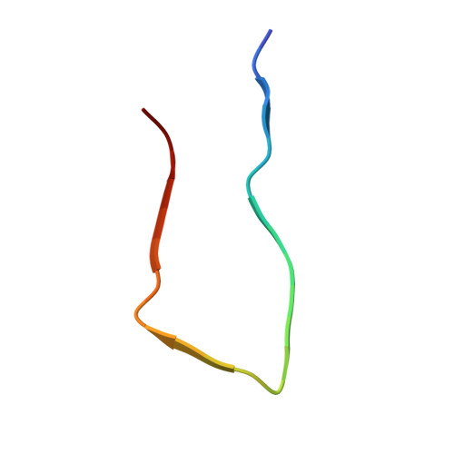
    Fibrils of the hamster prion peptide (sHaPrP, sequence 108-144) were prepared in an acidic solution, and their structure was solved by cryogenic electron microscopy with a resolution of 2.23 Å based on the gold-standard Fourier shell correlation (FSC) curve. The fibril has a novel architecture that has never been found in other amyloid fibrils. Each fibril is assembled by four protofilaments (PFs) and has an ordered water channel in the center. Each protofilament contains three β-strands (125-130, 133-135, and 138-141) arranged in an "R"-shaped construct. The structural data indicate that these three β-strand segments are the most amyloidogenic region of the prion peptide/protein and might be the site of nucleation during fibrillization under conditions without denaturants.


  • Organizational Affiliation

    Institute of Biological Chemistry, Academia Sinica, No. 128, Section 2, Academia Road, Nankang, Taipei 115, Taiwan.


Macromolecules

Find similar proteins by:  Sequence   |   3D Structure  

Entity ID: 1
MoleculeChains Sequence LengthOrganismDetailsImage
Major prion protein
A,
B,
C,
D,
E,
F,
G,
H,
I,
J,
K,
L,
M,
N,
O,
P,
Q,
R,
S,
T,
U,
V,
W [auth X],
X [auth Y]
25Mesocricetus auratusMutation(s): 0 
UniProt
Find proteins for P04273 (Mesocricetus auratus)
Explore P04273 
Go to UniProtKB:  P04273
Entity Groups  
Sequence Clusters30% Identity50% Identity70% Identity90% Identity95% Identity100% Identity
UniProt GroupP04273
Sequence Annotations
Expand
  • Reference Sequence
Experimental Data & Validation

Experimental Data

  • Method: ELECTRON MICROSCOPY
  • Resolution: 2.20 Å
  • Aggregation State: FILAMENT 
  • Reconstruction Method: HELICAL 
EM Software:
TaskSoftware PackageVersion
RECONSTRUCTIONRELION3.1
MODEL REFINEMENTPHENIX

Structure Validation

View Full Validation Report


Entry History & Funding Information

Deposition Data


Funding OrganizationLocationGrant Number
Academia Sinica (Taiwan)TaiwanAS-CDA-110-L03
Ministry of Science and Technology (MoST, Taiwan)TaiwanMOST 105-2119-M-001-028
Ministry of Science and Technology (MoST, Taiwan)Taiwan106-2923-M-001-003-MY3
Ministry of Science and Technology (MoST, Taiwan)Taiwan109-2113-M-001-001
Ministry of Science and Technology (MoST, Taiwan)TaiwanMOST 109-2311-B-001-022

Revision History  (Full details and data files)

  • Version 1.0: 2022-07-27
    Type: Initial release
  • Version 1.1: 2022-08-03
    Changes: Database references
  • Version 1.2: 2022-08-17
    Changes: Database references