


Crystal structure of Escherichia coli Adenine Phosphoribosyltransferase (APRT) in complex with Adenine
Yadav, P., Kushwaha, G.S., Bhavesh, N.S.To be published.
Experimental Data Snapshot
Starting Model: experimental
View more details
Entity ID: 1 | |||||
|---|---|---|---|---|---|
| Molecule | Chains | Sequence Length | Organism | Details | Image |
| Adenine phosphoribosyltransferase | 186 | Escherichia coli K-12 | Mutation(s): 0 Gene Names: apt, b0469, JW0458 EC: 2.4.2.7 | 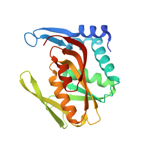 | |
UniProt | |||||
Entity Groups | |||||
| Sequence Clusters | 30% Identity50% Identity70% Identity90% Identity95% Identity100% Identity | ||||
| UniProt Group | P69503 | ||||
Sequence AnnotationsExpand | |||||
Reference Sequence | |||||
| Ligands 5 Unique | |||||
|---|---|---|---|---|---|
| ID | Chains | Name / Formula / InChI Key | 2D Diagram | 3D Interactions | |
| ADE (Subject of Investigation/LOI) Download:Ideal Coordinates CCD File | C [auth A], J [auth B] | ADENINE C5 H5 N5 GFFGJBXGBJISGV-UHFFFAOYSA-N |  | ||
| EDO Download:Ideal Coordinates CCD File | K [auth B] | 1,2-ETHANEDIOL C2 H6 O2 LYCAIKOWRPUZTN-UHFFFAOYSA-N |  | ||
| ACT Download:Ideal Coordinates CCD File | D [auth A], E [auth A] | ACETATE ION C2 H3 O2 QTBSBXVTEAMEQO-UHFFFAOYSA-M |  | ||
| CL Download:Ideal Coordinates CCD File | F [auth A], G [auth A], H [auth A], L [auth B], M [auth B] | CHLORIDE ION Cl VEXZGXHMUGYJMC-UHFFFAOYSA-M |  | ||
| NA Download:Ideal Coordinates CCD File | I [auth A], N [auth B] | SODIUM ION Na FKNQFGJONOIPTF-UHFFFAOYSA-N |  | ||
| Length ( Å ) | Angle ( ˚ ) |
|---|---|
| a = 44.656 | α = 90 |
| b = 87.073 | β = 111.2 |
| c = 47.83 | γ = 90 |
| Software Name | Purpose |
|---|---|
| REFMAC | refinement |
| autoPROC | data reduction |
| Aimless | data scaling |
| MOLREP | phasing |
| Funding Organization | Location | Grant Number |
|---|---|---|
| Indian Council of Medical Research | India | BMI/11(45)/2020 |


