7Y7J | pdb_00007y7j

SARS-CoV-2 S trimer in complex with 1F Fab


Experimental Data Snapshot

  • Method: ELECTRON MICROSCOPY
  • Resolution: 3.80 Å
  • Aggregation State: PARTICLE 
  • Reconstruction Method: SINGLE PARTICLE 

wwPDB Validation 3D Report Full Report

Validation slider image for 7Y7J

This is version 1.1 of the entry. See complete history

Literature

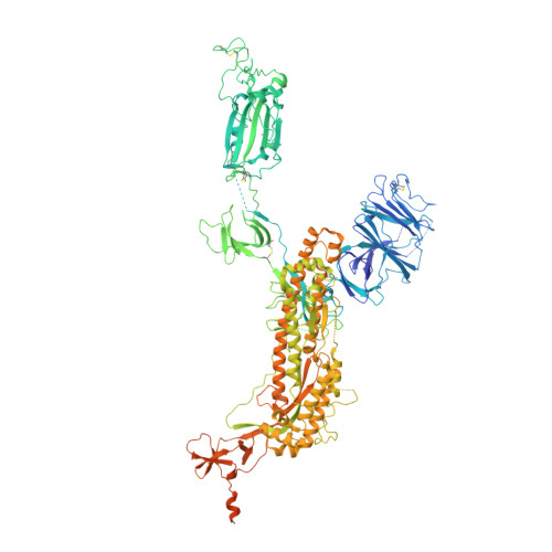
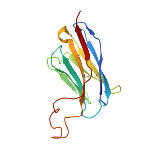
A core epitope targeting antibody of SARS-CoV-2.

Zhao, S.Liu, F.Qiu, S.Lan, Q.Wu, Y.Xu, W.Ke, J.Yang, J.Liu, X.Wang, K.Guo, H.Xia, S.Zhang, F.Wang, J.Hu, X.Lu, L.Jiang, S.Zhao, S.Liu, L.Xie, Y.Yang, X.Wang, H.Zhong, G.

(2023) Protein Cell 14: 74-78

  • DOI: https://doi.org/10.1093/procel/pwac042
  • Primary Citation Related Structures: 
    7Y7J, 7Y7K

  • Organizational Affiliation
    • iHuman Institute, ShanghaiTech University, Shanghai 201210, China.

Macromolecule Content 

  • Total Structure Weight: 465.32 kDa 
  • Atom Count: 23,742 
  • Modeled Residue Count: 3,191 
  • Deposited Residue Count: 4,107 
  • Unique protein chains: 3

Macromolecules

Find similar proteins by:|  3D Structure
Entity ID: 1
MoleculeChains  Sequence LengthOrganismDetailsImage
Spike glycoprotein
A, B, C
1,288Severe acute respiratory syndrome coronavirus 2Mutation(s): 5 
Gene Names: S2
UniProt
Find proteins for P0DTC2 (Severe acute respiratory syndrome coronavirus 2)
Explore P0DTC2 
Go to UniProtKB:  P0DTC2
Entity Groups
Sequence Clusters30% Identity50% Identity70% Identity90% Identity95% Identity100% Identity
UniProt GroupP0DTC2
Glycosylation
Glycosylation Sites: 15Go to GlyGen: P0DTC2-1
Sequence Annotations
Expand
Reference Sequence
Find similar proteins by:|  3D Structure
Entity ID: 2
MoleculeChains  Sequence LengthOrganismDetailsImage
1F VHD [auth E]132Homo sapiensMutation(s): 0 
Find similar proteins by:|  3D Structure
Entity ID: 3
MoleculeChains  Sequence LengthOrganismDetailsImage
1F VLE [auth D]111Homo sapiensMutation(s): 0 

Small Molecules

Ligands 1 Unique
IDChains Name / Formula / InChI Key2D Diagram3D Interactions
NAG

Query on NAG


Download:Ideal Coordinates CCD File
AA [auth A]
BA [auth A]
CA [auth B]
DA [auth B]
EA [auth B]
AA [auth A],
BA [auth A],
CA [auth B],
DA [auth B],
EA [auth B],
FA [auth B],
GA [auth B],
HA [auth B],
IA [auth B],
JA [auth B],
KA [auth B],
LA [auth C],
MA [auth C],
NA [auth C],
OA [auth C],
PA [auth C],
QA [auth C],
RA [auth C],
SA [auth C],
TA [auth C],
U [auth A],
V [auth A],
W [auth A],
X [auth A],
Y [auth A],
Z [auth A]
2-acetamido-2-deoxy-beta-D-glucopyranose
C8 H15 N O6
OVRNDRQMDRJTHS-FMDGEEDCSA-N

Experimental Data & Validation

Experimental Data

  • Method: ELECTRON MICROSCOPY
  • Resolution: 3.80 Å
  • Aggregation State: PARTICLE 
  • Reconstruction Method: SINGLE PARTICLE 

Structure Validation

View Full Validation Report


Entry History 

& Funding Information

Deposition Data


Funding OrganizationLocationGrant Number
National Natural Science Foundation of China (NSFC)China--

Revision History  (Full details and data files)

  • Version 1.0: 2023-02-22
    Type: Initial release
  • Version 1.1: 2024-11-13
    Changes: Data collection, Refinement description, Structure summary