7Y3W | pdb_00007y3w

Crystal structure of an questin oxidase (BTG13) from Cercospora sp. JNU001


Experimental Data Snapshot

  • Method: X-RAY DIFFRACTION
  • Resolution: 1.90 Å
  • R-Value Free: 
    0.182 (Depositor), 0.161 (DCC) 
  • R-Value Work: 
    0.150 (Depositor) 
  • R-Value Observed: 
    0.151 (Depositor) 

wwPDB Validation 3D Report Full Report

Validation slider image for 7Y3W

This is version 1.0 of the entry. See complete history

Literature

Discovery of the Biosynthetic Pathway of Beticolin 1 Reveals a Novel Non-Heme Iron-Dependent Oxygenase for Anthraquinone Ring Cleavage.

Hou, X.Xu, H.Deng, Z.Yan, Y.Yuan, Z.Liu, X.Su, Z.Yang, S.Zhang, Y.Rao, Y.

(2022) Angew Chem Int Ed Engl 61: e202208772-e202208772

  • DOI: https://doi.org/10.1002/anie.202208772
  • Primary Citation Related Structures: 
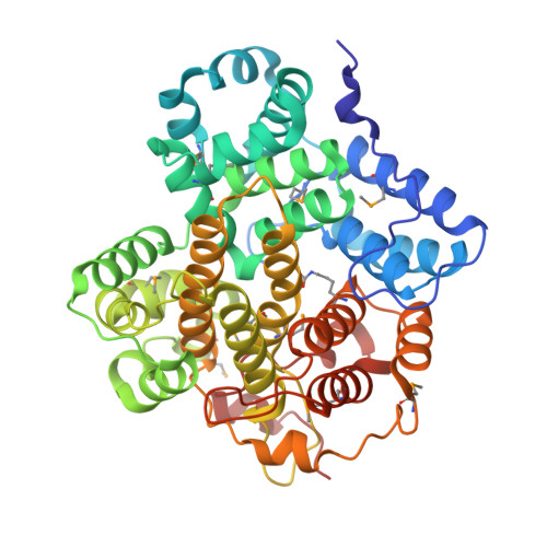
    7Y3W, 7Y3X, 7Y3Y

  • PubMed Abstract: 

    This study used light-mediated comparative transcriptomics to identify the biosynthetic gene cluster of beticolin 1 in Cercospora. It contains an anthraquinone moiety and an unusual halogenated xanthone moiety connected by a bicyclo[3.2.2]nonane. During elucidation of the biosynthetic pathway of beticolin 1, a novel non-heme iron oxygenase BTG13 responsible for anthraquinone ring cleavage was discovered. More importantly, the discovery of non-heme iron oxygenase BTG13 is well supported by experimental evidence: (i) crystal structure and the inductively coupled plasma mass spectrometry revealed that its reactive site is built by an atypical iron ion coordination, where the iron ion is uncommonly coordinated by four histidine residues, an unusual carboxylated-lysine (Kcx377) and water; (ii) Kcx377 is mediated by His58 and Thr299 to modulate the catalytic activity of BTG13. Therefore, we believed this study updates our knowledge of metalloenzymes.


  • Organizational Affiliation
    • Key Laboratory of Carbohydrate Chemistry and Biotechnology, Ministry of Education, School of Biotechnology, Jiangnan University, Wuxi, 214122, P. R. China.

Macromolecule Content 

  • Total Structure Weight: 101.14 kDa 
  • Atom Count: 7,818 
  • Modeled Residue Count: 880 
  • Deposited Residue Count: 888 
  • Unique protein chains: 1

Macromolecules

Find similar proteins by:|  3D Structure
Entity ID: 1
MoleculeChains  Sequence LengthOrganismDetailsImage
questin oxidase
A, B
444Cercospora sojinaMutation(s): 0 

Small Molecules

Ligands 2 Unique
IDChains Name / Formula / InChI Key2D Diagram3D Interactions
GOL

Query on GOL


Download:Ideal Coordinates CCD File
D [auth A]
E [auth A]
F [auth A]
G [auth A]
H [auth A]
D [auth A],
E [auth A],
F [auth A],
G [auth A],
H [auth A],
J [auth B],
K [auth B],
L [auth B],
M [auth B],
N [auth B],
O [auth B]
GLYCEROL
C3 H8 O3
PEDCQBHIVMGVHV-UHFFFAOYSA-N
FE
(Subject of Investigation/LOI)

Query on FE


Download:Ideal Coordinates CCD File
C [auth A],
I [auth B]
FE (III) ION
Fe
VTLYFUHAOXGGBS-UHFFFAOYSA-N
Modified Residues  2 Unique
IDChains TypeFormula2D DiagramParent
KCX
Query on KCX
A, B
L-PEPTIDE LINKINGC7 H14 N2 O4LYS
MSE
Query on MSE
A, B
L-PEPTIDE LINKINGC5 H11 N O2 SeMET

Experimental Data & Validation

Experimental Data

  • Method: X-RAY DIFFRACTION
  • Resolution: 1.90 Å
  • R-Value Free:  0.182 (Depositor), 0.161 (DCC) 
  • R-Value Work:  0.150 (Depositor) 
  • R-Value Observed: 0.151 (Depositor) 
Space Group: P 21 21 21
Unit Cell:
Length ( Å )Angle ( ˚ )
a = 78.781α = 90
b = 99.618β = 90
c = 130.638γ = 90
Software Package:
Software NamePurpose
REFMACrefinement
HKL-2000data scaling
PDB_EXTRACTdata extraction
HKL-2000data reduction
AutoSolphasing

Structure Validation

View Full Validation Report


Entry History 

& Funding Information

Deposition Data


Funding OrganizationLocationGrant Number
Other privateChina2018YFA0901700
Other privateChinaJUSRP12015
Other privateChina2020M671329
Other privateChina2020Z383

Revision History  (Full details and data files)

  • Version 1.0: 2022-09-21
    Type: Initial release