Arabidopsis Sec14 proteins (SFH5 and SFH7) mediate interorganelle transport of phosphatidic acid and regulate chloroplast development.
Yao, H.Y., Lu, Y.Q., Yang, X.L., Wang, X.Q., Luo, Z., Lin, D.L., Wu, J.W., Xue, H.W.(2023) Proc Natl Acad Sci U S A 120: e2221637120-e2221637120
- PubMed: 36716376 Search on PubMedSearch on PubMed Central
- DOI: https://doi.org/10.1073/pnas.2221637120
- Primary Citation Related Structures:
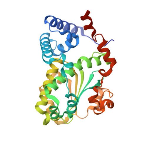
7Y10, 7Y11 - PubMed Abstract:
Lipids establish the specialized thylakoid membrane of chloroplast in eukaryotic photosynthetic organisms, while the molecular basis of lipid transfer from other organelles to chloroplast remains further elucidation. Here we revealed the structural basis of Arabidopsis Sec14 homology proteins AtSFH5 and AtSFH7 in transferring phosphatidic acid (PA) from endoplasmic reticulum (ER) to chloroplast, and whose function in regulating the lipid composition of chloroplast and thylakoid development. AtSFH5 and AtSFH7 localize at both ER and chloroplast, whose deficiency resulted in an abnormal chloroplast structure and a decreased thickness of stacked thylakoid membranes. We demonstrated that AtSFH5, but not yeast and human Sec14 proteins, could specifically recognize and transfer PA in vitro. Crystal structures of the AtSFH5-Sec14 domain in complex with L-α-phosphatidic acid (L-α-PA) and 1,2-dipalmitoyl-sn-glycero-3-phosphate (DPPA) revealed that two PA ligands nestled in the central cavity with different configurations, elucidating the specific binding mode of PA to AtSFH5, different from the reported phosphatidylethanolamine (PE)/phosphatidylcholine (PC)/phosphatidylinositol (PI) binding modes. Quantitative lipidomic analysis of chloroplast lipids showed that PA and monogalactosyldiacylglycerol (MGDG), particularly the C18 fatty acids at sn -2 position in MGDG were significantly decreased, indicating a disrupted ER-to-plastid (chloroplast) lipid transfer, under deficiency of AtSFH5 and AtSFH7. Our studies identified the role and elucidated the structural basis of plant SFH proteins in transferring PA between organelles, and suggested a model for ER-chloroplast interorganelle phospholipid transport from inherent ER to chloroplast derived from endosymbiosis of a cyanobacteriumproviding a mechanism involved in the adaptive evolution of cellular plastids.
- State Key Laboratory of Genetic Engineering, School of Life Sciences, Fudan University, Shanghai 200438, China.
Organizational Affiliation: