


Pre- and post-fusion class III glycoprotein structures reveal viral fusion mechanism
Shan, Y.Y., Zhang, M.F., Pei, D.Q.To be published.
Experimental Data Snapshot
wwPDB Validation 3D Report Full Report

Entity ID: 1 | |||||
|---|---|---|---|---|---|
| Molecule | Chains | Sequence Length | Organism | Details | Image |
| Envelope glycoprotein | 520 | Thogotovirus dhoriense | Mutation(s): 3 Gene Names: P4 | 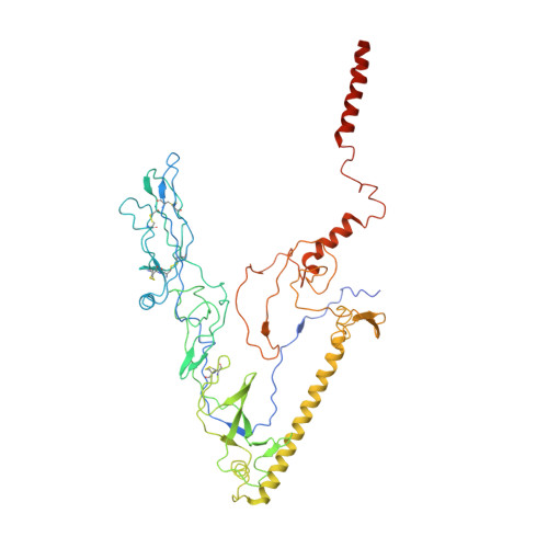 | |
UniProt | |||||
Entity Groups | |||||
| Sequence Clusters | 30% Identity50% Identity70% Identity90% Identity95% Identity100% Identity | ||||
| UniProt Group | P27427 | ||||
Sequence AnnotationsExpand | |||||
Reference Sequence | |||||
| Funding Organization | Location | Grant Number |
|---|---|---|
| National Natural Science Foundation of China (NSFC) | China | 2017YFA0504100 |


