7XV0

Crystal structure of RPA70N-BLMp1 fusion


Experimental Data Snapshot

  • Method: X-RAY DIFFRACTION
  • Resolution: 1.50 Å
  • R-Value Free: 0.200 
  • R-Value Work: 0.169 
  • R-Value Observed: 0.170 

Starting Model: experimental
View more details

wwPDB Validation   3D Report Full Report


This is version 1.3 of the entry. See complete history


Literature

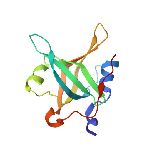
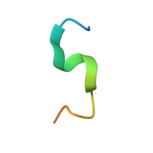
Structural characterization of human RPA70N association with DNA damage response proteins.

Wu, Y.Fu, W.Zang, N.Zhou, C.

(2023) Elife 12

  • DOI: https://doi.org/10.7554/eLife.81639
  • Primary Citation of Related Structures:  
    7XUT, 7XUV, 7XUW, 7XV0, 7XV1, 7XV4, 8JZV, 8JZY, 8K00

  • PubMed Abstract: 

    The heterotrimeric Replication protein A (RPA) is the ubiquitous eukaryotic single-stranded DNA (ssDNA) binding protein and participates in nearly all aspects of DNA metabolism, especially DNA damage response. The N-terminal OB domain of the RPA70 subunit (RPA70N) is a major protein-protein interaction element for RPA and binds to more than 20 partner proteins. Previous crystallography studies of RPA70N with p53, DNA2 and PrimPol fragments revealed that RPA70N binds to amphipathic peptides that mimic ssDNA. NMR chemical-shift studies also provided valuable information on the interaction of RPA70N residues with target sequences. However, it is still unclear how RPA70N recognizes and distinguishes such a diverse group of target proteins. Here, we present high-resolution crystal structures of RPA70N in complex with peptides from eight DNA damage response proteins. The structures show that, in addition to the ssDNA mimicry mode of interaction, RPA70N employs multiple ways to bind its partners. Our results advance the mechanistic understanding of RPA70N-mediated recruitment of DNA damage response proteins.


  • Organizational Affiliation

    School of Public Health & Sir Run Run Shaw Hospital, Zhejiang University School of Medicine, Zhejiang, China.


Macromolecules
Find similar proteins by:  (by identity cutoff)  |  3D Structure
Entity ID: 1
MoleculeChains Sequence LengthOrganismDetailsImage
Replication protein A 70 kDa DNA-binding subunit126Homo sapiensMutation(s): 0 
Gene Names: RPA1REPA1RPA70
UniProt & NIH Common Fund Data Resources
Find proteins for P27694 (Homo sapiens)
Explore P27694 
Go to UniProtKB:  P27694
PHAROS:  P27694
GTEx:  ENSG00000132383 
Entity Groups  
Sequence Clusters30% Identity50% Identity70% Identity90% Identity95% Identity100% Identity
UniProt GroupP27694
Sequence Annotations
Expand
  • Reference Sequence

Find similar proteins by:  Sequence   |   3D Structure  

Entity ID: 2
MoleculeChains Sequence LengthOrganismDetailsImage
Bloom syndrome protein20Homo sapiensMutation(s): 0 
Gene Names: BLMRECQ2RECQL3
EC: 3.6.4.12
UniProt & NIH Common Fund Data Resources
Find proteins for P54132 (Homo sapiens)
Explore P54132 
Go to UniProtKB:  P54132
PHAROS:  P54132
GTEx:  ENSG00000197299 
Entity Groups  
Sequence Clusters30% Identity50% Identity70% Identity90% Identity95% Identity100% Identity
UniProt GroupP54132
Sequence Annotations
Expand
  • Reference Sequence
Experimental Data & Validation

Experimental Data

  • Method: X-RAY DIFFRACTION
  • Resolution: 1.50 Å
  • R-Value Free: 0.200 
  • R-Value Work: 0.169 
  • R-Value Observed: 0.170 
  • Space Group: P 21 21 21
Unit Cell:
Length ( Å )Angle ( ˚ )
a = 38.088α = 90
b = 53.644β = 90
c = 54.501γ = 90
Software Package:
Software NamePurpose
PHENIXrefinement
PDB_EXTRACTdata extraction
XDSdata reduction
Aimlessdata scaling
PHASERphasing

Structure Validation

View Full Validation Report


Entry History & Funding Information

Deposition Data


Funding OrganizationLocationGrant Number
Ministry of Education (MoE, China)China--

Revision History  (Full details and data files)

  • Version 1.0: 2023-06-14
    Type: Initial release
  • Version 1.1: 2023-09-13
    Changes: Data collection, Database references
  • Version 1.2: 2023-11-29
    Changes: Refinement description
  • Version 1.3: 2024-05-08
    Changes: Database references