7XOT | pdb_00007xot

Crystal structure of Keap1_6k

  • Classification: PROTEIN BINDING
  • Organism(s): Homo sapiens
  • Expression System: Escherichia coli
  • Mutation(s): No 

  • Deposited: 2022-05-01 Released: 2023-05-03 
  • Deposition Author(s): Xu, K.
  • Funding Organization(s): Ministry of Science and Technology (MoST, China), National Natural Science Foundation of China (NSFC)

Experimental Data Snapshot

  • Method: X-RAY DIFFRACTION
  • Resolution: 2.70 Å
  • R-Value Free: 
    0.237 (Depositor), 0.240 (DCC) 
  • R-Value Work: 
    0.176 (Depositor), 0.180 (DCC) 
  • R-Value Observed: 
    0.182 (Depositor) 

Starting Model: experimental
View more details

wwPDB Validation   3D Report Full Report


Ligand Structure Quality Assessment 

Created with Raphaël 2.3.0Worse 01 BetterLigand structure goodness of fit to experimental dataBest fitted HB6Click on this verticalbar to view details

This is version 1.2 of the entry. See complete history


Literature

Crystal structure of Keap1_6k

Xu, K.

To be published.

Macromolecules
Find similar proteins by:  (by identity cutoff)  |  3D Structure
Entity ID: 1
MoleculeChains Sequence LengthOrganismDetailsImage
Kelch-like ECH-associated protein 1292Homo sapiensMutation(s): 0 
Gene Names: KEAP1
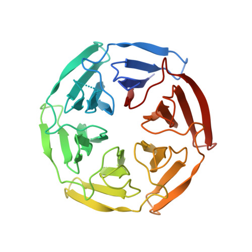
UniProt & NIH Common Fund Data Resources
Find proteins for Q14145 (Homo sapiens)
Explore Q14145 
Go to UniProtKB:  Q14145
PHAROS:  Q14145
GTEx:  ENSG00000079999 
Entity Groups  
Sequence Clusters30% Identity50% Identity70% Identity90% Identity95% Identity100% Identity
UniProt GroupQ14145
Sequence Annotations
Expand
  • Reference Sequence
Small Molecules
Ligands 1 Unique
IDChains Name / Formula / InChI Key2D Diagram3D Interactions
HB6 (Subject of Investigation/LOI)
Query on HB6

Download Ideal Coordinates CCD File 
B [auth A]2-[[4-[(2-azanyl-2-oxidanylidene-ethyl)-(4-methoxyphenyl)sulfonyl-amino]naphthalen-1-yl]-[4-(2-diethoxyphosphorylethanoylamino)phenyl]sulfonyl-amino]ethanamide
C33 H38 N5 O11 P S2
NQQGETZCYZQFPL-UHFFFAOYSA-N
Experimental Data & Validation

Experimental Data

  • Method: X-RAY DIFFRACTION
  • Resolution: 2.70 Å
  • R-Value Free:  0.237 (Depositor), 0.240 (DCC) 
  • R-Value Work:  0.176 (Depositor), 0.180 (DCC) 
  • R-Value Observed: 0.182 (Depositor) 
Space Group: C 1 2 1
Unit Cell:
Length ( Å )Angle ( ˚ )
a = 126.771α = 90
b = 76.309β = 105.622
c = 48.248γ = 90
Software Package:
Software NamePurpose
XDSdata reduction
XDSdata scaling
Cootmodel building
PHENIXphasing
PHENIXrefinement

Structure Validation

View Full Validation Report


Ligand Structure Quality Assessment 

Created with Raphaël 2.3.0Worse 01 BetterLigand structure goodness of fit to experimental dataBest fitted HB6Click on this verticalbar to view details

Entry History & Funding Information

Deposition Data

  • Released Date: 2023-05-03 
  • Deposition Author(s): Xu, K.

Funding OrganizationLocationGrant Number
Ministry of Science and Technology (MoST, China)China--
National Natural Science Foundation of China (NSFC)China--

Revision History  (Full details and data files)

  • Version 1.0: 2023-05-03
    Type: Initial release
  • Version 1.1: 2023-11-29
    Changes: Data collection, Refinement description
  • Version 1.2: 2024-10-16
    Changes: Structure summary