


Crystal Structure of Alpha-1,3-mannosyltransferase MNT2 from Saccharomyces cerevisiae, Mn/GDP-mannose form
Hira, D., Kadooka, C., Oka, T.To be published.
Experimental Data Snapshot
Starting Model: in silico
View more details
Entity ID: 1 | |||||
|---|---|---|---|---|---|
| Molecule | Chains | Sequence Length | Organism | Details | Image |
| Alpha-1,3-mannosyltransferase MNT2 | 548 | Saccharomyces cerevisiae W303 | Mutation(s): 0 Gene Names: MNT2, YGL257C, NRD558 EC: 2.4.1 | 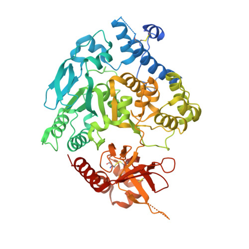 | |
UniProt | |||||
Entity Groups | |||||
| Sequence Clusters | 30% Identity50% Identity70% Identity90% Identity95% Identity100% Identity | ||||
| UniProt Group | P53059 | ||||
Sequence AnnotationsExpand | |||||
Reference Sequence | |||||
| Ligands 4 Unique | |||||
|---|---|---|---|---|---|
| ID | Chains | Name / Formula / InChI Key | 2D Diagram | 3D Interactions | |
| GDD (Subject of Investigation/LOI) Download:Ideal Coordinates CCD File | CA [auth D], F [auth A], N [auth B], U [auth C] | GUANOSINE-5'-DIPHOSPHATE-ALPHA-D-MANNOSE C16 H25 N5 O16 P2 MVMSCBBUIHUTGJ-GDJBGNAASA-N |  | ||
| GOL Download:Ideal Coordinates CCD File | DA [auth D] EA [auth D] FA [auth D] G [auth A] GA [auth D] | GLYCEROL C3 H8 O3 PEDCQBHIVMGVHV-UHFFFAOYSA-N |  | ||
| MN (Subject of Investigation/LOI) Download:Ideal Coordinates CCD File | BA [auth D], E [auth A], M [auth B], T [auth C] | MANGANESE (II) ION Mn WAEMQWOKJMHJLA-UHFFFAOYSA-N |  | ||
| NA Download:Ideal Coordinates CCD File | AA [auth C], HA [auth D], L [auth A], S [auth B], Z [auth C] | SODIUM ION Na FKNQFGJONOIPTF-UHFFFAOYSA-N |  | ||
| Length ( Å ) | Angle ( ˚ ) |
|---|---|
| a = 130.849 | α = 90 |
| b = 131.189 | β = 90 |
| c = 169.552 | γ = 90 |
| Software Name | Purpose |
|---|---|
| REFMAC | refinement |
| XDS | data reduction |
| XSCALE | data scaling |
| PHASER | phasing |
| Funding Organization | Location | Grant Number |
|---|---|---|
| Not funded | -- |


