7XID | pdb_00007xid

S-ECD (Omicron) in complex with PD of ACE2


Experimental Data Snapshot

  • Method: ELECTRON MICROSCOPY
  • Resolution: 3.30 Å
  • Aggregation State: PARTICLE 
  • Reconstruction Method: SINGLE PARTICLE 

wwPDB Validation 3D Report Full Report

Validation slider image for 7XID

This is version 1.2 of the entry. See complete history

Literature

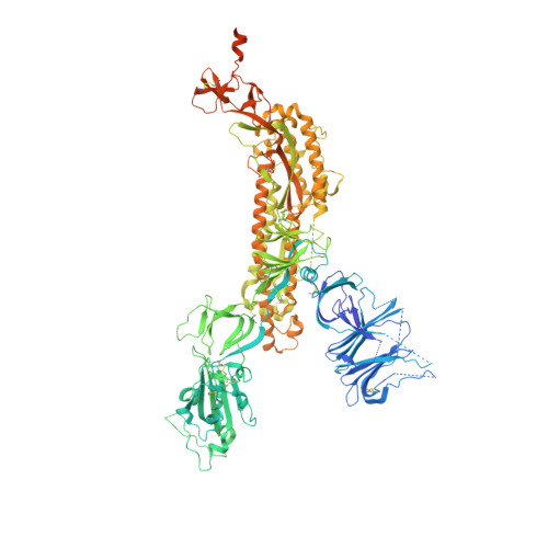
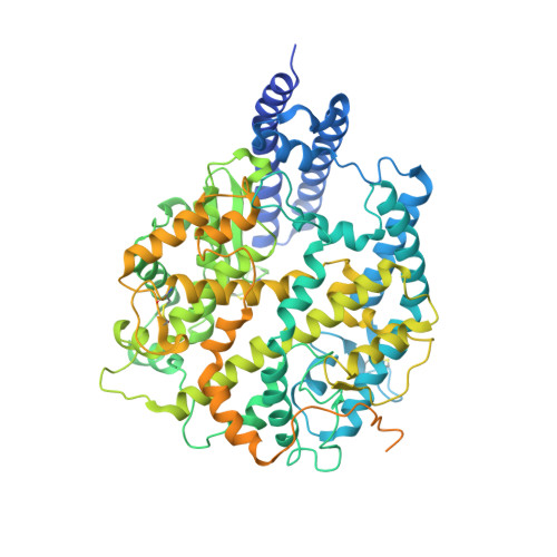
Structural and functional analysis of an inter-Spike bivalent neutralizing antibody against SARS-CoV-2 variants.

Li, Y.Fan, Q.Zhou, B.Shen, Y.Zhang, Y.Cheng, L.Qi, F.Song, S.Guo, Y.Yan, R.Ju, B.Zhang, Z.

(2022) iScience 25: 104431-104431

  • DOI: https://doi.org/10.1016/j.isci.2022.104431
  • Primary Citation Related Structures: 
    7XIC, 7XID

  • PubMed Abstract: 

    The different variants of severe acute respiratory syndrome coronavirus 2 (SARS-CoV-2) have attracted most public concern because they caused "wave and wave" COVID-19 pandemic. The initial step of viral infection is mediated by the SARS-CoV-2 Spike (S) protein, which mediates the receptor recognition and membrane fusion between virus and host cells. Neutralizing antibodies (nAbs) targeting the S protein of SARS-CoV-2 have become promising candidates for clinical intervention strategy, while multiple studies have shown that different variants have enhanced infectivity and antibody resistance. Here, we explore the structure and function of STS165, a broadly inter-Spike bivalent nAb against SARS-CoV-2 variants and even SARS-CoV, contributing to further understanding of the working mechanism of nAbs.


  • Organizational Affiliation
    • Center for Infectious Disease Research, Westlake Laboratory of Life Sciences and Biomedicine, Key Laboratory of Structural Biology of Zhejiang Province, School of Life Sciences, Westlake University, Zhejiang Province, Hangzhou 310024, China.

Macromolecule Content 

  • Total Structure Weight: 630.54 kDa 
  • Atom Count: 34,256 
  • Modeled Residue Count: 4,153 
  • Deposited Residue Count: 5,435 
  • Unique protein chains: 2

Macromolecules

Find similar proteins by:|  3D Structure
Entity ID: 1
MoleculeChains  Sequence LengthOrganismDetailsImage
Spike glycoprotein
A, B, C
1,267Severe acute respiratory syndrome coronavirus 2Mutation(s): 39 
Gene Names: S2
UniProt
Find proteins for P0DTC2 (Severe acute respiratory syndrome coronavirus 2)
Explore P0DTC2 
Go to UniProtKB:  P0DTC2
Entity Groups
Sequence Clusters30% Identity50% Identity70% Identity90% Identity95% Identity100% Identity
UniProt GroupP0DTC2
Glycosylation
Glycosylation Sites: 16Go to GlyGen: P0DTC2-1
Sequence Annotations
Expand
Reference Sequence
Find similar proteins by:|  3D Structure
Entity ID: 2
MoleculeChains  Sequence LengthOrganismDetailsImage
Angiotensin-converting enzyme 2
D, E
817Homo sapiensMutation(s): 0 
Gene Names: ACE2UNQ868/PRO1885
EC: 3.4.17.23 (PDB Primary Data), 3.4.17 (PDB Primary Data)
UniProt & NIH Common Fund Data Resources
Find proteins for Q9BYF1 (Homo sapiens)
Explore Q9BYF1 
Go to UniProtKB:  Q9BYF1
PHAROS:  Q9BYF1
GTEx:  ENSG00000130234 
Entity Groups
Sequence Clusters30% Identity50% Identity70% Identity90% Identity95% Identity100% Identity
UniProt GroupQ9BYF1
Glycosylation
Glycosylation Sites: 4Go to GlyGen: Q9BYF1-1
Sequence Annotations
Expand
Reference Sequence

Small Molecules

Ligands 1 Unique
IDChains Name / Formula / InChI Key2D Diagram3D Interactions
NAG
(Subject of Investigation/LOI)

Query on NAG


Download:Ideal Coordinates CCD File
AB [auth C]
BB [auth C]
CA [auth A]
CB [auth C]
DA [auth A]
AB [auth C],
BB [auth C],
CA [auth A],
CB [auth C],
DA [auth A],
DB [auth C],
EA [auth A],
EB [auth C],
FA [auth A],
FB [auth C],
GA [auth A],
GB [auth C],
HA [auth A],
HB [auth D],
IA [auth A],
IB [auth D],
JA [auth A],
JB [auth D],
KA [auth A],
KB [auth D],
LA [auth A],
LB [auth D],
MA [auth A],
MB [auth D],
NA [auth B],
NB [auth D],
OA [auth B],
OB [auth E],
PA [auth B],
PB [auth E],
QA [auth B],
QB [auth E],
RA [auth B],
RB [auth E],
SA [auth B],
SB [auth E],
TA [auth B],
TB [auth E],
UA [auth B],
UB [auth E],
VA [auth B],
WA [auth B],
XA [auth B],
YA [auth C],
ZA [auth C]
2-acetamido-2-deoxy-beta-D-glucopyranose
C8 H15 N O6
OVRNDRQMDRJTHS-FMDGEEDCSA-N

Experimental Data & Validation

Experimental Data

  • Method: ELECTRON MICROSCOPY
  • Resolution: 3.30 Å
  • Aggregation State: PARTICLE 
  • Reconstruction Method: SINGLE PARTICLE 
EM Software:
TaskSoftware PackageVersion
RECONSTRUCTIONRELION3.0.6

Structure Validation

View Full Validation Report


Entry History 

& Funding Information

Deposition Data


Funding OrganizationLocationGrant Number
National Natural Science Foundation of China (NSFC)China92169204

Revision History  (Full details and data files)

  • Version 1.0: 2022-06-15
    Type: Initial release
  • Version 1.1: 2022-06-22
    Changes: Database references
  • Version 1.2: 2024-10-30
    Changes: Data collection, Structure summary