7XG8 | pdb_00007xg8

Crystal structure of PstS protein from cyanophage P-SSM2


Experimental Data Snapshot

  • Method: X-RAY DIFFRACTION
  • Resolution: 2.25 Å
  • R-Value Free: 
    0.279 (Depositor), 0.282 (DCC) 
  • R-Value Work: 
    0.227 (Depositor), 0.234 (DCC) 
  • R-Value Observed: 
    0.229 (Depositor) 

Starting Model: experimental
View more details

wwPDB Validation 3D Report Full Report

Validation slider image for 7XG8

Ligand Structure Quality Assessment 


This is version 1.2 of the entry. See complete history

Literature

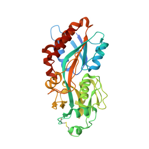
Biochemical and structural characterization of the cyanophage-encoded phosphate-binding protein: implications for enhanced phosphate uptake of infected cyanobacteria

Zhao, F.Lin, X.Cai, K.Jiang, Y.Ni, T.Chen, Y.Feng, J.Dang, S.Zhou, C.Z.Zeng, Q.

(2022) Environ Microbiol 24: 3037-3050

Macromolecule Content 

  • Total Structure Weight: 33.91 kDa 
  • Atom Count: 2,248 
  • Modeled Residue Count: 298 
  • Deposited Residue Count: 321 
  • Unique protein chains: 1

Macromolecules

Find similar proteins by:|  3D Structure
Entity ID: 1
MoleculeChains  Sequence LengthOrganismDetailsImage
ABC transporter, substrate binding protein, phosphate321Prochlorococcus phage P-SSM2Mutation(s): 0 
Gene Names: pstSPCMG_00251PSSM2_248
UniProt
Find proteins for Q58MA7 (Prochlorococcus phage P-SSM2)
Explore Q58MA7 
Go to UniProtKB:  Q58MA7
Entity Groups
Sequence Clusters30% Identity50% Identity70% Identity90% Identity95% Identity100% Identity
UniProt GroupQ58MA7
Sequence Annotations
Expand
Reference Sequence

Small Molecules

Ligands 1 Unique
IDChains Name / Formula / InChI Key2D Diagram3D Interactions
PO4
(Subject of Investigation/LOI)

Query on PO4


Download:Ideal Coordinates CCD File
B [auth A]PHOSPHATE ION
O4 P
NBIIXXVUZAFLBC-UHFFFAOYSA-K

Experimental Data & Validation

Experimental Data

  • Method: X-RAY DIFFRACTION
  • Resolution: 2.25 Å
  • R-Value Free:  0.279 (Depositor), 0.282 (DCC) 
  • R-Value Work:  0.227 (Depositor), 0.234 (DCC) 
  • R-Value Observed: 0.229 (Depositor) 
Space Group: P 21 21 21
Unit Cell:
Length ( Å )Angle ( ˚ )
a = 38.774α = 90
b = 63.525β = 90
c = 108.083γ = 90
Software Package:
Software NamePurpose
REFMACrefinement
PDB_EXTRACTdata extraction
HKL-2000data reduction
HKL-2000data scaling
MOLREPphasing

Structure Validation

View Full Validation Report


Ligand Structure Quality Assessment 


Entry History 

& Funding Information

Deposition Data


Funding OrganizationLocationGrant Number
Not funded--

Revision History  (Full details and data files)

  • Version 1.0: 2023-02-08
    Type: Initial release
  • Version 1.1: 2023-11-29
    Changes: Data collection, Refinement description
  • Version 1.2: 2024-11-06
    Changes: Structure summary