7XC0 | pdb_00007xc0

Crystal structure of Human RPTPH


Experimental Data Snapshot

  • Method: X-RAY DIFFRACTION
  • Resolution: 1.56 Å
  • R-Value Free: 
    0.207 (Depositor), 0.207 (DCC) 
  • R-Value Work: 
    0.178 (Depositor), 0.178 (DCC) 
  • R-Value Observed: 
    0.180 (Depositor) 

Starting Model: experimental
View more details

wwPDB Validation 3D Report Full Report

Validation slider image for 7XC0

This is version 1.2 of the entry. See complete history

Literature

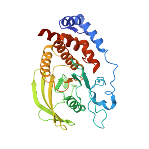
Crystal structure of the catalytic domain of human RPTPH.

Kim, M.Ryu, S.E.

(2022) Acta Crystallogr F Struct Biol Commun 78: 265-269

  • DOI: https://doi.org/10.1107/S2053230X22006173
  • Primary Citation Related Structures: 
    7XC0

  • PubMed Abstract: 

    Receptor-type protein tyrosine phosphatases (RPTPs) receive extracellular stimuli and transfer them into cells. They regulate cell growth, differentiation and death via specific signals. They have also been implicated in cancer, diabetes and neurological diseases. RPTPH, a member of the type 3 RPTP (R3-PTP) family, is an important regulator of colorectal cancer and hepatic carcinoma. Despite its importance in drug development, the structure of RPTPH has not yet been resolved. Here, the crystal structure of the catalytic domain of RPTPH was determined at 1.56 Å resolution. Despite similarities to other R3-PTPs in its overall structure, RPTPH exhibited differences in its loop regions and side-chain conformations. Compared with other R3-PTPs, RPTPH has unique side chains near its active site that may confer specificity for inhibitor binding. Therefore, detailed information on the structure of RPTPH provides clues for the development of specific inhibitors.


  • Organizational Affiliation
    • Department of Bioengineering, College of Engineering, Hanyang University, Seoul 04673, Republic of Korea.

Macromolecule Content 

  • Total Structure Weight: 33.32 kDa 
  • Atom Count: 2,512 
  • Modeled Residue Count: 287 
  • Deposited Residue Count: 291 
  • Unique protein chains: 1

Macromolecules

Find similar proteins by:|  3D Structure
Entity ID: 1
MoleculeChains  Sequence LengthOrganismDetailsImage
Receptor-type tyrosine-protein phosphatase H291Homo sapiensMutation(s): 2 
Gene Names: PTPRHSAP1
EC: 3.1.3.48
UniProt & NIH Common Fund Data Resources
Find proteins for Q9HD43 (Homo sapiens)
Explore Q9HD43 
Go to UniProtKB:  Q9HD43
PHAROS:  Q9HD43
GTEx:  ENSG00000080031 
Entity Groups
Sequence Clusters30% Identity50% Identity70% Identity90% Identity95% Identity100% Identity
UniProt GroupQ9HD43
Sequence Annotations
Expand
Reference Sequence

Small Molecules

Ligands 1 Unique
IDChains Name / Formula / InChI Key2D Diagram3D Interactions
PO4

Query on PO4


Download:Ideal Coordinates CCD File
B [auth A]PHOSPHATE ION
O4 P
NBIIXXVUZAFLBC-UHFFFAOYSA-K

Experimental Data & Validation

Experimental Data

  • Method: X-RAY DIFFRACTION
  • Resolution: 1.56 Å
  • R-Value Free:  0.207 (Depositor), 0.207 (DCC) 
  • R-Value Work:  0.178 (Depositor), 0.178 (DCC) 
  • R-Value Observed: 0.180 (Depositor) 
Space Group: P 32
Unit Cell:
Length ( Å )Angle ( ˚ )
a = 56.46α = 90
b = 56.46β = 90
c = 80.447γ = 120
Software Package:
Software NamePurpose
PHENIXrefinement
HKL-2000data reduction
HKL-2000data scaling
PHENIXphasing

Structure Validation

View Full Validation Report


Entry History 

& Funding Information

Deposition Data


Funding OrganizationLocationGrant Number
National Research Foundation (NRF, Korea)Korea, Republic Of--

Revision History  (Full details and data files)

  • Version 1.0: 2022-07-06
    Type: Initial release
  • Version 1.1: 2022-07-20
    Changes: Database references
  • Version 1.2: 2023-11-29
    Changes: Data collection, Refinement description