7XAO

Crystal structure of thioredoxin 1


Experimental Data Snapshot

  • Method: X-RAY DIFFRACTION
  • Resolution: 1.45 Å
  • R-Value Free: 0.283 
  • R-Value Work: 0.246 
  • R-Value Observed: 0.247 

wwPDB Validation   3D Report Full Report


This is version 1.1 of the entry. See complete history


Literature

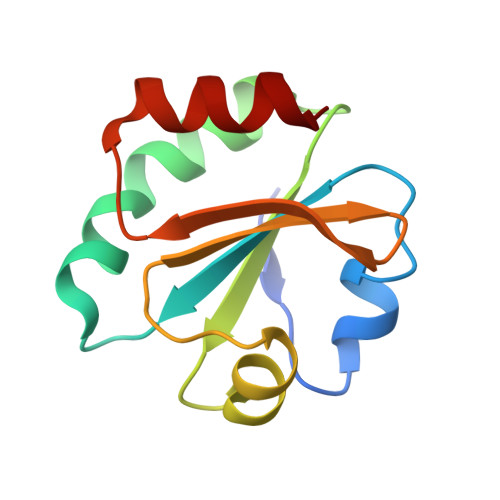
High-resolution crystal structure of Acinetobacter baumannii thioredoxin 1.

Chang, Y.J.Park, H.H.

(2022) Biochem Biophys Res Commun 608: 1-7

  • DOI: https://doi.org/10.1016/j.bbrc.2022.03.134
  • Primary Citation of Related Structures:  
    7XAO

  • PubMed Abstract: 

    Thioredoxin (Trx) is a central component of the redox control system that maintains the redox homeostasis critical for organism survival. Owing to its central role in survival, Trx is a prospective target for novel antimicrobial agents. Herein, we report a 1.45 Å high-resolution structure of Trx1 of Acinetobacter baumannii (abTrx1), an antibiotic-resistant pathogenic superbug. Although abTrx1 exhibited the canonical Trx fold, which consists of a four-stranded β-sheet surrounded by four α-helices, structural differences were detected in the loop forming the C-X-X-C redox center and the C-terminal. The unique CAPC sequence of the C-X-X-C motif in the abTrx1 redox center was characterized by mutagenesis. This study contributes to the field of drug designing against superbugs.


  • Organizational Affiliation

    College of Pharmacy, Chung-Ang University, Seoul, 06974, Republic of Korea; Department of Global Innovative Drugs, Graduate School of Chung-Ang University, Seoul, 06974, Republic of Korea.


Macromolecules
Find similar proteins by:  (by identity cutoff)  |  3D Structure
Entity ID: 1
MoleculeChains Sequence LengthOrganismDetailsImage
Thioredoxin
A, B
107Acinetobacter baumanniiMutation(s): 0 
Gene Names: trxAtrx
UniProt
Find proteins for V5VGM8 (Acinetobacter baumannii)
Explore V5VGM8 
Go to UniProtKB:  V5VGM8
Entity Groups  
Sequence Clusters30% Identity50% Identity70% Identity90% Identity95% Identity100% Identity
UniProt GroupV5VGM8
Sequence Annotations
Expand
  • Reference Sequence
Experimental Data & Validation

Experimental Data

  • Method: X-RAY DIFFRACTION
  • Resolution: 1.45 Å
  • R-Value Free: 0.283 
  • R-Value Work: 0.246 
  • R-Value Observed: 0.247 
  • Space Group: P 21 21 21
Unit Cell:
Length ( Å )Angle ( ˚ )
a = 31.24α = 90
b = 31.27β = 90
c = 215.78γ = 90
Software Package:
Software NamePurpose
PHENIXrefinement
PHENIXrefinement
PDB_EXTRACTdata extraction
XDSdata reduction
XDSdata scaling
PHENIXphasing

Structure Validation

View Full Validation Report


Entry History & Funding Information

Deposition Data


Funding OrganizationLocationGrant Number
National Research Foundation (NRF, Korea)Korea, Republic Of--

Revision History  (Full details and data files)

  • Version 1.0: 2022-06-01
    Type: Initial release
  • Version 1.1: 2023-11-29
    Changes: Data collection, Refinement description