7X7N | pdb_00007x7n

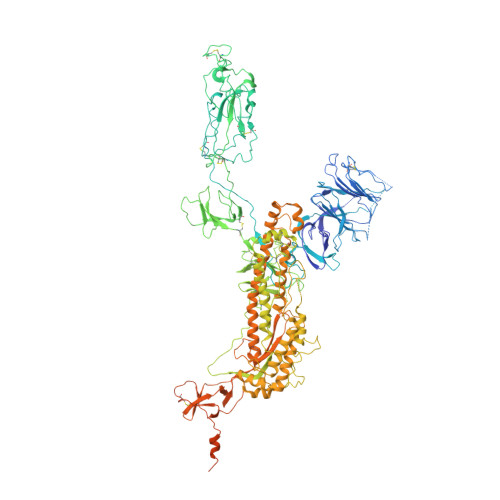
3D model of the 3-RBD up single trimeric spike protein of SARS-CoV2 in the presence of synthetic peptide SIH-5.


Experimental Data Snapshot

  • Method: ELECTRON MICROSCOPY
  • Resolution: 4.47 Å
  • Aggregation State: PARTICLE 
  • Reconstruction Method: SINGLE PARTICLE 

wwPDB Validation 3D Report Full Report

Validation slider image for 7X7N

This is version 1.2 of the entry. See complete history. 

Literature

A dimeric proteomimetic prevents SARS-CoV-2 infection by dimerizing the spike protein.

Khatri, B., Pramanick, I., Malladi, S.K., Rajmani, R.S., Kumar, S., Ghosh, P., Sengupta, N., Rahisuddin, R., Kumar, N., Kumaran, S., Ringe, R.P., Varadarajan, R., Dutta, S., Chatterjee, J.

(2022) Nat Chem Biol 18: 1046-1055

  • DOI: https://doi.org/10.1038/s41589-022-01060-0
  • Primary Citation Related Structures: 
    7X7N

  • PubMed Abstract: 

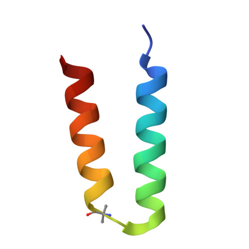
    Protein tertiary structure mimetics are valuable tools to target large protein-protein interaction interfaces. Here, we demonstrate a strategy for designing dimeric helix-hairpin motifs from a previously reported three-helix-bundle miniprotein that targets the receptor-binding domain (RBD) of severe acute respiratory syndrome-coronavirus-2 (SARS-CoV-2). Through truncation of the third helix and optimization of the interhelical loop residues of the miniprotein, we developed a thermostable dimeric helix-hairpin. The dimeric four-helix bundle competes with the human angiotensin-converting enzyme 2 (ACE2) in binding to RBD with 2:2 stoichiometry. Cryogenic-electron microscopy revealed the formation of dimeric spike ectodomain trimer by the four-helix bundle, where all the three RBDs from either spike protein are attached head-to-head in an open conformation, revealing a novel mechanism for virus neutralization. The proteomimetic protects hamsters from high dose viral challenge with replicative SARS-CoV-2 viruses, demonstrating the promise of this class of peptides that inhibit protein-protein interaction through target dimerization.


  • Organizational Affiliation: 
    • Molecular Biophysics Unit (MBU), Indian Institute of Science, Bangalore, India.

Macromolecule Content 

  • Total Structure Weight: 463.76 kDa 
  • Atom Count: 26,493 
  • Modeled Residue Count: 3,320 
  • Deposited Residue Count: 4,092 
  • Unique protein chains: 2

Macromolecules

Find similar proteins by:|  3D Structure
Entity ID: 1
MoleculeChains  Sequence LengthOrganismDetailsImage
Spike glycoproteinA [auth B],
B [auth C],
C [auth A]
1,288Severe acute respiratory syndrome coronavirus 2Mutation(s): 5 
Gene Names: S, 2
UniProt
Find proteins for P0DTC2 (Severe acute respiratory syndrome coronavirus 2)
Explore P0DTC2 
Go to UniProtKB:  P0DTC2
Entity Groups
Sequence Clusters30% Identity50% Identity70% Identity90% Identity95% Identity100% Identity
UniProt GroupP0DTC2
Glycosylation
Glycosylation Sites: 13Go to GlyGen: P0DTC2-1
Sequence Annotations
Expand
Reference Sequence
Find similar proteins by:|  3D Structure
Entity ID: 2
MoleculeChains  Sequence LengthOrganismDetailsImage
Synthetic peptide SIH-538synthetic constructMutation(s): 0 
Entity Groups
Sequence Clusters30% Identity50% Identity70% Identity90% Identity95% Identity100% Identity
Sequence Annotations
Expand
Reference Sequence

Small Molecules

Ligands 1 Unique
IDChains Name / Formula / InChI Key2D Diagram3D Interactions
NAG

Query on NAG


Download:Ideal Coordinates CCD File
AA [auth C]
BA [auth C]
CA [auth C]
DA [auth C]
EA [auth C]
AA [auth C],
BA [auth C],
CA [auth C],
DA [auth C],
EA [auth C],
FA [auth C],
GA [auth C],
HA [auth C],
IA [auth C],
J [auth B],
JA [auth A],
K [auth B],
KA [auth A],
L [auth B],
LA [auth A],
M [auth B],
MA [auth A],
N [auth B],
NA [auth A],
O [auth B],
OA [auth A],
P [auth B],
PA [auth A],
Q [auth B],
QA [auth A],
R [auth B],
RA [auth A],
S [auth B],
SA [auth A],
T [auth B],
TA [auth A],
U [auth B],
UA [auth A],
V [auth B],
VA [auth A],
W [auth C],
X [auth C],
Y [auth C],
Z [auth C]
2-acetamido-2-deoxy-beta-D-glucopyranose
C8 H15 N O6
OVRNDRQMDRJTHS-FMDGEEDCSA-N
Modified Residues  2 Unique
IDChains TypeFormula2D DiagramParent
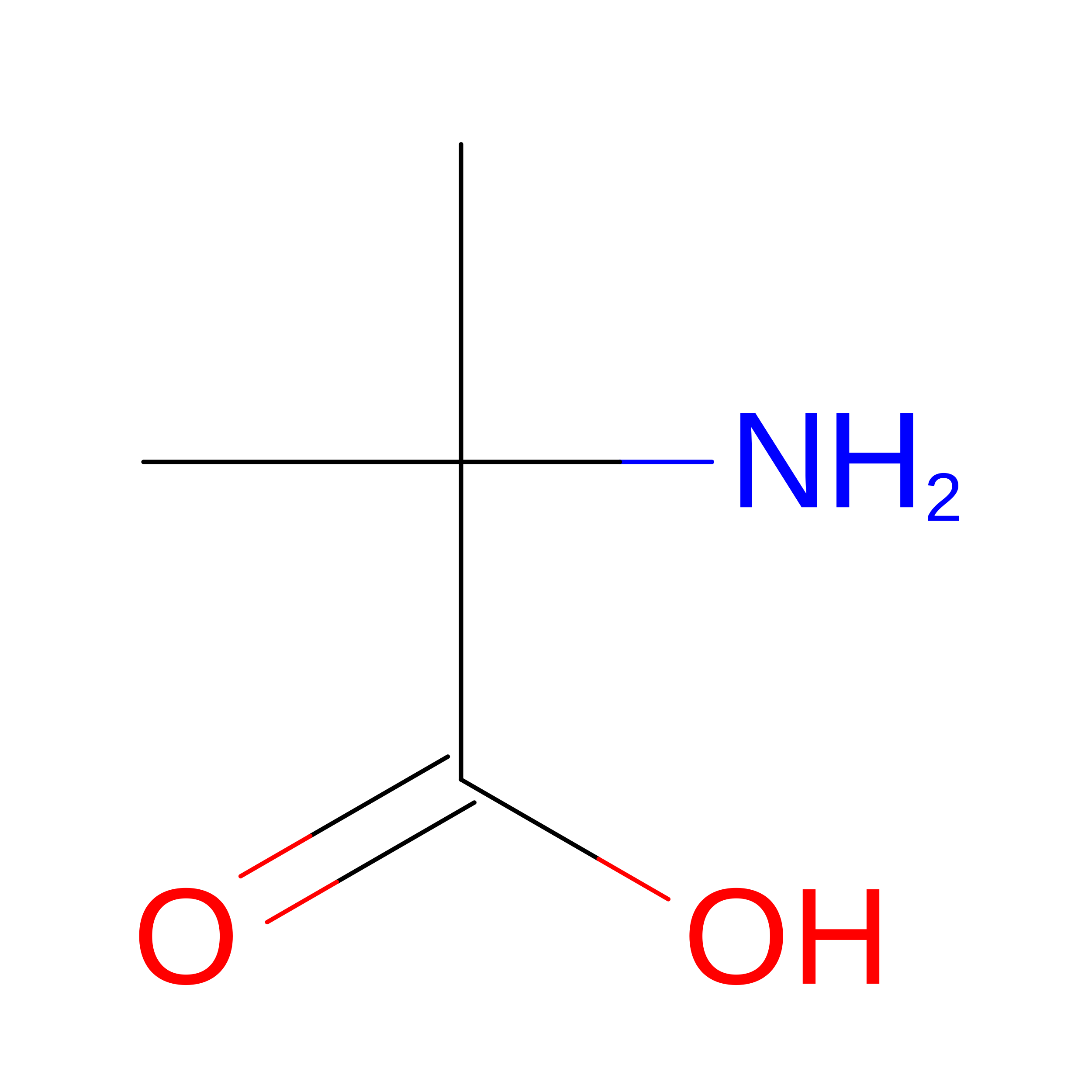
AIB
Query on AIB
D
E
F [auth H]
G [auth I]
H [auth F]
D,
E,
F [auth H],
G [auth I],
H [auth F],
I [auth G]
L-PEPTIDE LINKINGC4 H9 N O2ALA
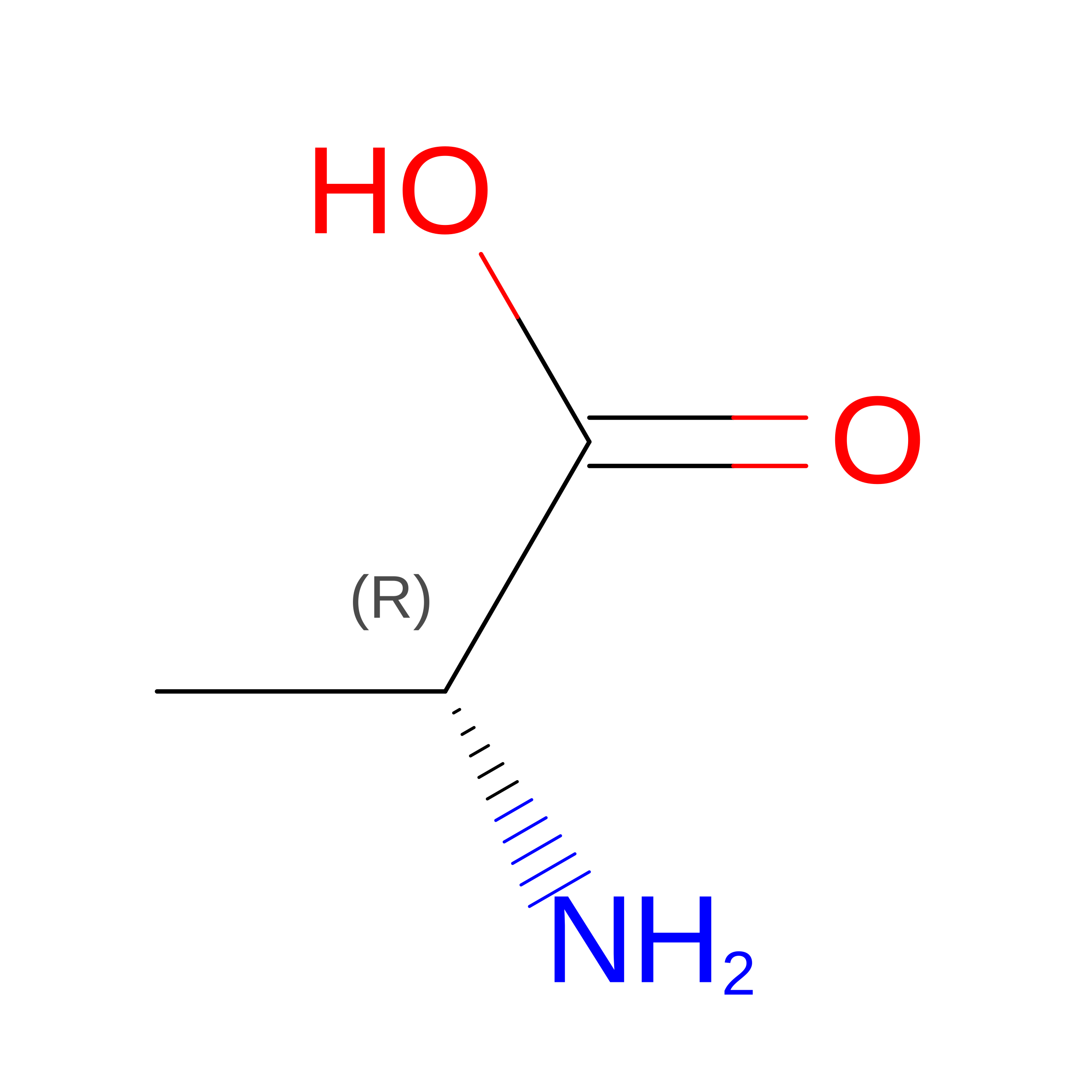
DAL
Query on DAL
D
E
F [auth H]
G [auth I]
H [auth F]
D,
E,
F [auth H],
G [auth I],
H [auth F],
I [auth G]
D-PEPTIDE LINKINGC3 H7 N O2

--

Experimental Data & Validation

Experimental Data

  • Method: ELECTRON MICROSCOPY
  • Resolution: 4.47 Å
  • Aggregation State: PARTICLE 
  • Reconstruction Method: SINGLE PARTICLE 
EM Software:
TaskSoftware PackageVersion
MODEL REFINEMENTPHENIX

Structure Validation

View Full Validation Report


Entry History 

& Funding Information

Deposition Data


Funding OrganizationLocationGrant Number
Department of Biotechnology (DBT, India)IndiaBT/INF/22/SP22844/2017
Department of Science & Technology (DST, India)IndiaSR/FST/LSII-039/2015

Revision History  (Full details and data files)

  • Version 1.0: 2022-04-27
    Type: Initial release
  • Version 1.1: 2022-11-16
    Changes: Database references
  • Version 1.2: 2025-07-02
    Changes: Data collection, Structure summary