


Crystal structure of human SIRT5 in complex with diazirine inhibitor 9
Li, G.-B., Deng, J.To be published.
Experimental Data Snapshot
Starting Model: experimental
View more details
Entity ID: 1 | |||||
|---|---|---|---|---|---|
| Molecule | Chains | Sequence Length | Organism | Details | Image |
| NAD-dependent protein deacylase sirtuin-5, mitochondrial | 277 | Homo sapiens | Mutation(s): 0 Gene Names: SIRT5, SIR2L5 EC: 2.3.1 | 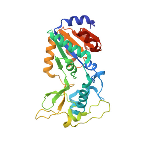 | |
UniProt & NIH Common Fund Data Resources | |||||
PHAROS: Q9NXA8 GTEx: ENSG00000124523 | |||||
Entity Groups | |||||
| Sequence Clusters | 30% Identity50% Identity70% Identity90% Identity95% Identity100% Identity | ||||
| UniProt Group | Q9NXA8 | ||||
Sequence AnnotationsExpand | |||||
Reference Sequence | |||||
| Ligands 3 Unique | |||||
|---|---|---|---|---|---|
| ID | Chains | Name / Formula / InChI Key | 2D Diagram | 3D Interactions | |
| 8VG (Subject of Investigation/LOI) Download:Ideal Coordinates CCD File | B [auth A] | 5-[[(5~{S})-6-[[(1~{S})-1-(4-hydroxyphenyl)-2-oxidanylidene-2-(prop-2-ynylamino)ethyl]amino]-6-oxidanylidene-5-[[4-[3-(trifluoromethyl)-1,2-diazirin-3-yl]phenyl]carbonylamino]hexyl]amino]-5-sulfanylidene-pentanoic acid C31 H33 F3 N6 O6 S BSPJSERNTAGWAV-OZXSUGGESA-N |  | ||
| PEG (Subject of Investigation/LOI) Download:Ideal Coordinates CCD File | C [auth A] | DI(HYDROXYETHYL)ETHER C4 H10 O3 MTHSVFCYNBDYFN-UHFFFAOYSA-N |  | ||
| ZN Download:Ideal Coordinates CCD File | D [auth A] | ZINC ION Zn PTFCDOFLOPIGGS-UHFFFAOYSA-N |  | ||
| Length ( Å ) | Angle ( ˚ ) |
|---|---|
| a = 41.332 | α = 90 |
| b = 65.034 | β = 93.58 |
| c = 55.658 | γ = 90 |
| Software Name | Purpose |
|---|---|
| HKL-2000 | data reduction |
| SCALEPACK | data scaling |
| PHASER | phasing |
| PHENIX | refinement |
| PDB_EXTRACT | data extraction |
| Funding Organization | Location | Grant Number |
|---|---|---|
| National Natural Science Foundation of China (NSFC) | China | 82122065, 82073698 |


