


Structure of tubulin H393D mutant from Odinarchaeota
Robinson, R.C., Ali, S., Narita, A.To be published.
Experimental Data Snapshot
wwPDB Validation 3D Report Full Report

Entity ID: 1 | |||||
|---|---|---|---|---|---|
| Molecule | Chains | Sequence Length | Organism | Details | Image |
| CBg-ParM triple mutant R204D, K230D and N234D | 290 | Clostridium botulinum | Mutation(s): 0 | 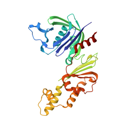 | |
| Length ( Å ) | Angle ( ˚ ) |
|---|---|
| a = 55.635 | α = 90 |
| b = 51.103 | β = 115.28 |
| c = 64.917 | γ = 90 |
| Software Name | Purpose |
|---|---|
| PHENIX | refinement |
| HKL-2000 | data reduction |
| HKL-2000 | data scaling |
| PHASER | phasing |
| Funding Organization | Location | Grant Number |
|---|---|---|
| Japan Society for the Promotion of Science (JSPS) | Japan | 18H02410 |
| Japan Society for the Promotion of Science (JSPS) | Japan | 21H02440 |
| Japan Science and Technology | Japan | JPMJCR19S5 |


