7X3A | pdb_00007x3a

NMR solution structure of the 1:1 complex of a pyridostatin (PDS) bound to a G-quadruplex MYT1L

  • Classification: DNA
  • Organism(s): Homo sapiens
  • Mutation(s): No 

  • Deposited: 2022-02-28 Released: 2022-06-08 
  • Deposition Author(s): Liu, L.-Y., Mao, Z.-W., Liu, W.
  • Funding Organization(s): National Natural Science Foundation of China (NSFC)

Experimental Data Snapshot

  • Method: SOLUTION NMR
  • Conformers Calculated: 100 
  • Conformers Submitted: 15 
  • Selection Criteria: structures with the lowest energy 

wwPDB Validation 3D Report Full Report

Validation slider image for 7X3A

This is version 1.3 of the entry. See complete history

Literature

Structural Basis of Pyridostatin and Its Derivatives Specifically Binding to G-Quadruplexes.

Liu, L.-Y.Ma, T.Z.Zeng, Y.L.Liu, W.Mao, Z.-W.

(2022) J Am Chem Soc 144: 11878-11887

  • DOI: https://doi.org/10.1021/jacs.2c04775
  • Primary Citation Related Structures: 
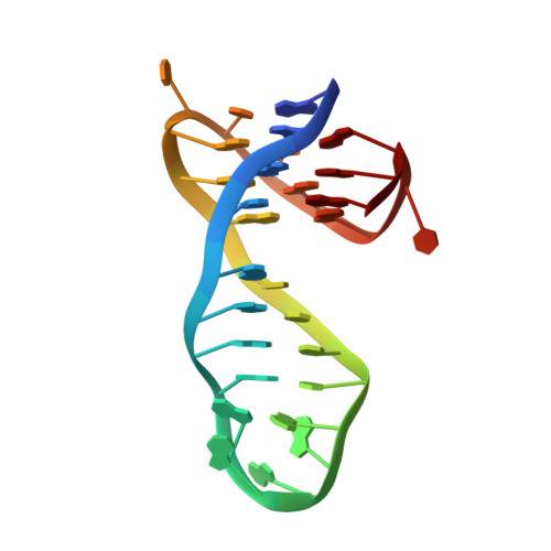
    7X2Z, 7X3A

  • PubMed Abstract: 

    The nucleic acid G-quadruplex (G4) has emerged as a promising therapeutic target for a variety of diseases such as cancer and neurodegenerative disease. Among small-molecule G4-binders, pyridostatin (PDS) and its derivatives ( e.g. , PyPDS) exhibit high specificity to G4s, but the structural basis for their specific recognition of G4s remains unknown. Here, we presented two solution structures of PyPDS and PDS with a quadruplex-duplex hybrid. The structures indicate that the rigid aromatic rings of PyPDS/PDS linked by flexible amide bonds match adaptively with G-tetrad planes, enhancing π-π stacking and achieving specific recognition of G4s. The aliphatic amine side chains of PyPDS/PDS adjust conformation to interact with the phosphate backbone via hydrogen bonding and electrostatic interactions, increasing affinity for G4s. Moreover, the N-H of PyPDS/PDS amide bonds interacts with two O 6 s of G-tetrad guanines via hydrogen bonding, achieving a further increase in affinity for G4s, which is different from most G4 ligands. Our findings reveal from structural perspectives that the rational assembly of rigid and flexible structural units in a ligand can synergistically improve the selectivity and affinity for G4s through spatial selective and adaptive matching.


  • Organizational Affiliation
    • MOE Key Laboratory of Bioinorganic and Synthetic Chemistry, School of Chemistry, Sun Yat-Sen University, Guangzhou 510275, P. R. China.

Macromolecule Content 

  • Total Structure Weight: 9.81 kDa 
  • Atom Count: 658 
  • Modeled Residue Count: 29 
  • Deposited Residue Count: 29 
  • Unique nucleic acid chains: 1

Macromolecules

Find similar nucleic acids by:  (by identity cutoff) 
Entity ID: 1
MoleculeChains LengthOrganismImage
G-quadruplex DNA MYT1L29Homo sapiens
Sequence Annotations
Expand
Reference Sequence

Small Molecules

Ligands 1 Unique
IDChains Name / Formula / InChI Key2D Diagram3D Interactions
7V8
(Subject of Investigation/LOI)

Query on 7V8


Download:Ideal Coordinates CCD File
B [auth A]4-(2-azanylethoxy)-N2,N6-bis[4-(2-azanylethoxy)quinolin-2-yl]pyridine-2,6-dicarboxamide
C31 H32 N8 O5
VGHSATQVJCTKEF-UHFFFAOYSA-N

Experimental Data & Validation

Experimental Data

  • Method: SOLUTION NMR
  • Conformers Calculated: 100 
  • Conformers Submitted: 15 
  • Selection Criteria: structures with the lowest energy 

Structure Validation

View Full Validation Report


Entry History 

& Funding Information

Deposition Data


Funding OrganizationLocationGrant Number
National Natural Science Foundation of China (NSFC)China--

Revision History  (Full details and data files)

  • Version 1.0: 2022-06-08
    Type: Initial release
  • Version 1.1: 2022-12-21
    Changes: Database references
  • Version 1.2: 2023-06-14
    Changes: Other
  • Version 1.3: 2024-05-15
    Changes: Data collection, Database references