7WYT | pdb_00007wyt

Crystal structures of Na+,K+-ATPase in complex with ouabain


Experimental Data Snapshot

  • Method: X-RAY DIFFRACTION
  • Resolution: 2.90 Å
  • R-Value Free: 
    0.307 (Depositor), 0.311 (DCC) 
  • R-Value Work: 
    0.270 (Depositor), 0.278 (DCC) 
  • R-Value Observed: 
    0.274 (Depositor) 

Starting Model: experimental
View more details

wwPDB Validation 3D Report Full Report

Validation slider image for 7WYT

Ligand Structure Quality Assessment 


This is version 1.1 of the entry. See complete history

Literature

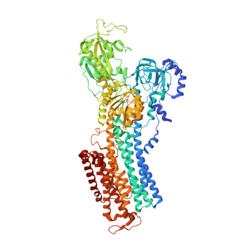
Cryoelectron microscopy of Na + ,K + -ATPase in the two E2P states with and without cardiotonic steroids.

Kanai, R.Cornelius, F.Vilsen, B.Toyoshima, C.

(2022) Proc Natl Acad Sci U S A 119: e2123226119-e2123226119

  • DOI: https://doi.org/10.1073/pnas.2123226119
  • Primary Citation Related Structures: 
    7WYS, 7WYT, 7WYU, 7WYV, 7WYW, 7WYX, 7WYY, 7WYZ, 7WZ0

  • PubMed Abstract: 

    Cryoelectron microscopy (cryo-EM) was applied to Na+,K+-ATPase (NKA) to determine the structures of two E2P states, one (E2PATP) formed by ATP and Mg2+ in the forward reaction, and the other (E2PPi) formed by inorganic phosphate (Pi) and Mg2+ in the backward reaction, with and without ouabain or istaroxime, representatives of classical and new-generation cardiotonic steroids (CTSs). These two E2P states exhibit different biochemical properties. In particular, K+-sensitive acceleration of the dephosphorylation reaction is not observed with E2PPi, attributed to the presence of a Mg2+ ion in the transmembrane cation binding sites. The cryo-EM structures of NKA demonstrate that the two E2P structures are nearly identical but Mg2+ in the transmembrane binding cavity is identified only in E2PPi, corroborating the idea that it should be denoted as E2PPi·Mg2+. We can now explain why the absence of transmembrane Mg2+ in E2PATP confers the K+ sensitivity in dephosphorylation. In addition, we show that ATP bridges the actuator (A) and nucleotide binding (N) domains, stabilizing the E2PATP state; CTS binding causes hardly any changes in the structure of NKA, both in E2PATP and E2PPi·Mg2+, indicating that the binding mechanism is conformational selection; and istaroxime binds to NKA, extending its aminoalkyloxime group deep into the cation binding site. This orientation is upside down compared to that of classical CTSs with respect to the steroid ring. Notably, mobile parts of NKA are resolved substantially better in the electron microscopy (EM) maps than in previous X-ray structures, including sugars sticking out from the β-subunit and many phospholipid molecules.


  • Organizational Affiliation
    • Institute for Quantitative Biosciences, The University of Tokyo, Bunkyo-ku, Tokyo 113-0032, Japan.

Macromolecule Content 

  • Total Structure Weight: 325.02 kDa 
  • Atom Count: 21,354 
  • Modeled Residue Count: 2,632 
  • Deposited Residue Count: 2,768 
  • Unique protein chains: 3

Macromolecules

Find similar proteins by:|  3D Structure
Entity ID: 1
MoleculeChains  Sequence LengthOrganismDetailsImage
Sodium/potassium-transporting ATPase subunit alpha-1A,
D [auth C]
1,016Sus scrofaMutation(s): 0 
EC: 7.2.2.13
Membrane Entity: Yes 
UniProt
Find proteins for P05024 (Sus scrofa)
Explore P05024 
Go to UniProtKB:  P05024
Entity Groups
Sequence Clusters30% Identity50% Identity70% Identity90% Identity95% Identity100% Identity
UniProt GroupP05024
Sequence Annotations
Expand
Reference Sequence
Find similar proteins by:|  3D Structure
Entity ID: 2
MoleculeChains  Sequence LengthOrganismDetailsImage
Sodium/potassium-transporting ATPase subunit beta-1B,
E [auth D]
303Sus scrofaMutation(s): 0 
Membrane Entity: Yes 
UniProt
Find proteins for P05027 (Sus scrofa)
Explore P05027 
Go to UniProtKB:  P05027
Entity Groups
Sequence Clusters30% Identity50% Identity70% Identity90% Identity95% Identity100% Identity
UniProt GroupP05027
Glycosylation
Glycosylation Sites: 3
Sequence Annotations
Expand
Reference Sequence
Find similar proteins by:|  3D Structure
Entity ID: 3
MoleculeChains  Sequence LengthOrganismDetailsImage
FXYD domain-containing ion transport regulatorC [auth G],
F [auth E]
65Sus scrofaMutation(s): 0 
Membrane Entity: Yes 
UniProt
Find proteins for Q58K79 (Sus scrofa)
Explore Q58K79 
Go to UniProtKB:  Q58K79
Entity Groups
Sequence Clusters30% Identity50% Identity70% Identity90% Identity95% Identity100% Identity
UniProt GroupQ58K79
Sequence Annotations
Expand
Reference Sequence

Oligosaccharides

Help  
Entity ID: 4
MoleculeChains Length2D Diagram GlycosylationD Interactions
2-acetamido-2-deoxy-beta-D-glucopyranose-(1-4)-2-acetamido-2-deoxy-beta-D-glucopyranoseG [auth F],
H,
I,
J
2N-Glycosylation
Glycosylation Resources
GlyTouCan: G42666HT
GlyCosmos: G42666HT
GlyGen: G42666HT

Small Molecules

Ligands 6 Unique
IDChains Name / Formula / InChI Key2D Diagram3D Interactions
PCW

Query on PCW


Download:Ideal Coordinates CCD File
FA [auth C]
GA [auth C]
HA [auth C]
IA [auth C]
O [auth A]
FA [auth C],
GA [auth C],
HA [auth C],
IA [auth C],
O [auth A],
P [auth A],
Q [auth A],
R [auth A],
S [auth A],
T [auth A],
U [auth A],
V [auth A],
W [auth A]
1,2-DIOLEOYL-SN-GLYCERO-3-PHOSPHOCHOLINE
C44 H85 N O8 P
SNKAWJBJQDLSFF-NVKMUCNASA-O
OBN
(Subject of Investigation/LOI)

Query on OBN


Download:Ideal Coordinates CCD File
JA [auth C],
X [auth A]
OUABAIN
C29 H44 O12
LPMXVESGRSUGHW-HBYQJFLCSA-N
CLR

Query on CLR


Download:Ideal Coordinates CCD File
DA [auth C],
EA [auth C],
LA [auth D],
N [auth A],
Z [auth G]
CHOLESTEROL
C27 H46 O
HVYWMOMLDIMFJA-DPAQBDIFSA-N
NAG

Query on NAG


Download:Ideal Coordinates CCD File
KA [auth D],
Y [auth B]
2-acetamido-2-deoxy-beta-D-glucopyranose
C8 H15 N O6
OVRNDRQMDRJTHS-FMDGEEDCSA-N
MG

Query on MG


Download:Ideal Coordinates CCD File
AA [auth C],
CA [auth C],
K [auth A],
M [auth A]
MAGNESIUM ION
Mg
JLVVSXFLKOJNIY-UHFFFAOYSA-N
NA

Query on NA


Download:Ideal Coordinates CCD File
BA [auth C],
L [auth A]
SODIUM ION
Na
FKNQFGJONOIPTF-UHFFFAOYSA-N
Modified Residues  1 Unique
IDChains TypeFormula2D DiagramParent
PHD
Query on PHD
A,
D [auth C]
L-PEPTIDE LINKINGC4 H8 N O7 PASP

Experimental Data & Validation

Experimental Data

  • Method: X-RAY DIFFRACTION
  • Resolution: 2.90 Å
  • R-Value Free:  0.307 (Depositor), 0.311 (DCC) 
  • R-Value Work:  0.270 (Depositor), 0.278 (DCC) 
  • R-Value Observed: 0.274 (Depositor) 
Space Group: P 21 21 21
Unit Cell:
Length ( Å )Angle ( ˚ )
a = 115.621α = 90
b = 117.81β = 90
c = 493.203γ = 90
Software Package:
Software NamePurpose
HKL-2000data reduction
SCALEPACKdata scaling
MOLREPphasing
PHENIXrefinement

Structure Validation

View Full Validation Report


Ligand Structure Quality Assessment 


Entry History 

& Funding Information

Deposition Data


Funding OrganizationLocationGrant Number
Japan Society for the Promotion of Science (JSPS)Japan16K18500
Japan Society for the Promotion of Science (JSPS)Japan16H02499
Japan Society for the Promotion of Science (JSPS)Japan19H00975

Revision History  (Full details and data files)

  • Version 1.0: 2022-05-04
    Type: Initial release
  • Version 1.1: 2023-11-29
    Changes: Data collection, Refinement description