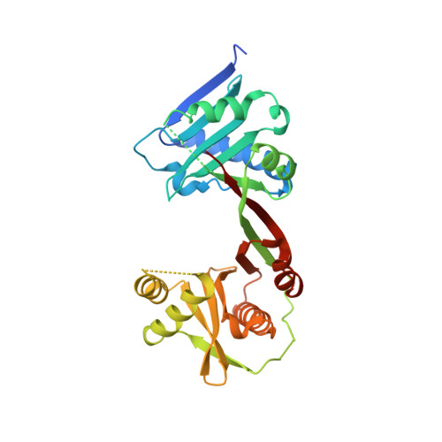
7WX6

A Legionella acetyltransferase VipF


Experimental Data Snapshot

  • Method: X-RAY DIFFRACTION
  • Resolution: 2.27 Å
  • R-Value Free: 0.253 
  • R-Value Work: 0.221 
  • R-Value Observed: 0.224 

Starting Model: experimental
View more details

wwPDB Validation   3D Report Full Report


Ligand Structure Quality Assessment 


This is version 1.1 of the entry. See complete history


Literature

Structural basis for the acetylation mechanism of the Legionella effector VipF.

Chen, T.T.Lin, Y.Zhang, S.Han, A.

(2022) Acta Crystallogr D Struct Biol 78: 1110-1119

  • DOI: https://doi.org/10.1107/S2059798322007318
  • Primary Citation of Related Structures:  
    7WX5, 7WX6, 7WX7

  • PubMed Abstract: 

    The pathogen Legionella pneumophila, which is the causative agent of Legionnaires' disease, secrets hundreds of effectors into host cells via its Dot/Icm secretion system to subvert host-cell pathways during pathogenesis. VipF, a conserved core effector among Legionella species, is a putative acetyltransferase, but its structure and catalytic mechanism remain unknown. Here, three crystal structures of VipF in complex with its cofactor acetyl-CoA and/or a substrate are reported. The two GNAT-like domains of VipF are connected as two wings by two β-strands to form a U-shape. Both domains bind acetyl-CoA or CoA, but only in the C-terminal domain does the molecule extend to the bottom of the U-shaped groove as required for an active transferase reaction; the molecule in the N-terminal domain folds back on itself. Interestingly, when chloramphenicol, a putative substrate, binds in the pocket of the central U-shaped groove adjacent to the N-terminal domain, VipF remains in an open conformation. Moreover, mutations in the central U-shaped groove, including Glu129 and Asp251, largely impaired the acetyltransferase activity of VipF, suggesting a unique enzymatic mechanism for the Legionella effector VipF.


  • Organizational Affiliation

    State Key Laboratory for Cellular Stress Biology, School of Life Sciences and Faculty of Medical Sciences, Xiamen University, Xiamen 361102, People's Republic of China.


Macromolecules
Find similar proteins by:  (by identity cutoff)  |  3D Structure
Entity ID: 1
MoleculeChains Sequence LengthOrganismDetailsImage
N-acetyltransferase305Legionella pneumophilaMutation(s): 0 
Gene Names: vipFC3927_15730DI026_06115
UniProt
Find proteins for Q5C8M4 (Legionella pneumophila)
Explore Q5C8M4 
Go to UniProtKB:  Q5C8M4
Entity Groups  
Sequence Clusters30% Identity50% Identity70% Identity90% Identity95% Identity100% Identity
UniProt GroupQ5C8M4
Sequence Annotations
Expand
  • Reference Sequence
Experimental Data & Validation

Experimental Data

  • Method: X-RAY DIFFRACTION
  • Resolution: 2.27 Å
  • R-Value Free: 0.253 
  • R-Value Work: 0.221 
  • R-Value Observed: 0.224 
  • Space Group: C 2 2 21
Unit Cell:
Length ( Å )Angle ( ˚ )
a = 72.047α = 90
b = 84.525β = 90
c = 105.547γ = 90
Software Package:
Software NamePurpose
PHENIXrefinement
HKL-2000data scaling
PDB_EXTRACTdata extraction
HKL-2000data reduction
PHENIXphasing

Structure Validation

View Full Validation Report


Ligand Structure Quality Assessment 


Entry History & Funding Information

Deposition Data


Funding OrganizationLocationGrant Number
National Natural Science Foundation of China (NSFC)China31770803
National Natural Science Foundation of China (NSFC)China81861138047

Revision History  (Full details and data files)

  • Version 1.0: 2022-09-14
    Type: Initial release
  • Version 1.1: 2023-11-29
    Changes: Data collection, Refinement description