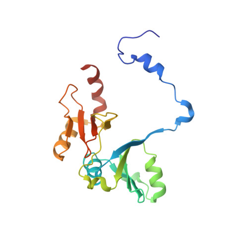
7WT0 | pdb_00007wt0

human glyoxalase I (with C-ter His tag) in complex with TLSC702

  • Classification: LYASE
  • Organism(s): Homo sapiens
  • Expression System: Escherichia coli
  • Mutation(s): No 

  • Deposited: 2022-02-03 Released: 2022-04-13 
  • Deposition Author(s): Usami, M., Yokoyama, H.
  • Funding Organization(s): Japan Society for the Promotion of Science (JSPS)

Experimental Data Snapshot

  • Method: X-RAY DIFFRACTION
  • Resolution: 2.00 Å
  • R-Value Free: 
    0.222 (Depositor), 0.230 (DCC) 
  • R-Value Work: 
    0.186 (Depositor), 0.200 (DCC) 
  • R-Value Observed: 
    0.190 (Depositor) 

Starting Model: experimental
View more details

wwPDB Validation   3D Report Full Report


Ligand Structure Quality Assessment 

Created with Raphaël 2.3.0Worse 01 BetterLigand structure goodness of fit to experimental dataBest fitted 5ZOClick on this verticalbar to view details

This is version 1.2 of the entry. See complete history


Literature

Crystal structures of human glyoxalase I and its complex with TLSC702 reveal inhibitor binding mode and substrate preference.

Usami, M.Ando, K.Shibuya, A.Takasawa, R.Yokoyama, H.

(2022) FEBS Lett 596: 1458-1467

  • DOI: https://doi.org/10.1002/1873-3468.14344
  • Primary Citation of Related Structures:  
    7WSZ, 7WT0, 7WT1, 7WT2

  • PubMed Abstract: 

    Human glyoxalase I (hGLO I) is an enzyme for detoxification of methylglyoxal (MG) and has been considered an attractive target for the development of new anticancer drugs. In our previous report, the GLO I inhibitor TLSC702 induced apoptosis in tumor cells. Here, we determined the crystal structures of hGLO I and its complex with TLSC702. In the complex, the carboxyl O atom of TLSC702 is coordinated to Zn 2+ , and TLSC702 mainly shows van der Waals interaction with hydrophobic residues. In the inhibitor-unbound structure, glycerol, which has similar functional groups to MG, was bound to Zn 2+ , indicating that GLO I can easily bind to MG. This study provides a structural basis to develop better anticancer drugs.


  • Organizational Affiliation

    Faculty of Pharmaceutical Sciences, Tokyo University of Science, Noda, Japan.


Macromolecules
Find similar proteins by:  (by identity cutoff)  |  3D Structure
Entity ID: 1
MoleculeChains Sequence LengthOrganismDetailsImage
Lactoylglutathione lyase
A, B
192Homo sapiensMutation(s): 0 
Gene Names: GLO1
EC: 4.4.1.5
UniProt & NIH Common Fund Data Resources
Find proteins for Q04760 (Homo sapiens)
Explore Q04760 
Go to UniProtKB:  Q04760
PHAROS:  Q04760
GTEx:  ENSG00000124767 
Entity Groups  
Sequence Clusters30% Identity50% Identity70% Identity90% Identity95% Identity100% Identity
UniProt GroupQ04760
Sequence Annotations
Expand
  • Reference Sequence
Experimental Data & Validation

Experimental Data

  • Method: X-RAY DIFFRACTION
  • Resolution: 2.00 Å
  • R-Value Free:  0.222 (Depositor), 0.230 (DCC) 
  • R-Value Work:  0.186 (Depositor), 0.200 (DCC) 
  • R-Value Observed: 0.190 (Depositor) 
Space Group: P 1 21 1
Unit Cell:
Length ( Å )Angle ( ˚ )
a = 53.689α = 90
b = 81.971β = 93.85
c = 46.927γ = 90
Software Package:
Software NamePurpose
REFMACrefinement
PDB_EXTRACTdata extraction
XDSdata reduction
SCALAdata scaling
MOLREPphasing

Structure Validation

View Full Validation Report


Ligand Structure Quality Assessment 

Created with Raphaël 2.3.0Worse 01 BetterLigand structure goodness of fit to experimental dataBest fitted 5ZOClick on this verticalbar to view details

Entry History & Funding Information

Deposition Data


Funding OrganizationLocationGrant Number
Japan Society for the Promotion of Science (JSPS)Japan17K07316

Revision History  (Full details and data files)

  • Version 1.0: 2022-04-13
    Type: Initial release
  • Version 1.1: 2022-06-29
    Changes: Database references
  • Version 1.2: 2023-11-29
    Changes: Data collection, Refinement description